7JSN

Structure of the Visual Signaling Complex between Transducin and Phosphodiesterase 6

  • Classification: SIGNALING PROTEIN
  • Organism(s): Bos taurus
  • Expression System: Escherichia coli
  • Mutation(s): Yes 
  • Membrane Protein: Yes  mpstruc

  • Deposited: 2020-08-15 Released: 2020-10-21 
  • Deposition Author(s): Gao, Y., Eskici, G., Ramachandran, S., Skiniotis, G., Cerione, R.A.
  • Funding Organization(s): National Institutes of Health/National Institute of General Medical Sciences (NIH/NIGMS), National Institutes of Health/National Cancer Institute (NIH/NCI), National Institutes of Health/National Institute of Neurological Disorders and Stroke (NIH/NINDS)

Experimental Data Snapshot

  • Method: ELECTRON MICROSCOPY
  • Resolution: 3.20 Å
  • Aggregation State: PARTICLE 
  • Reconstruction Method: SINGLE PARTICLE 

wwPDB Validation   3D Report Full Report


This is version 2.0 of the entry. See complete history


Literature

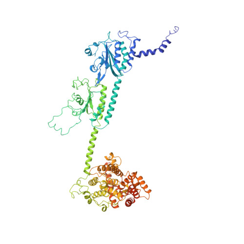
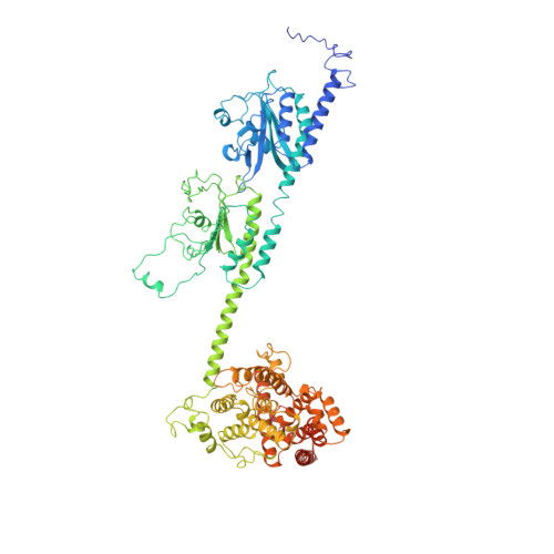
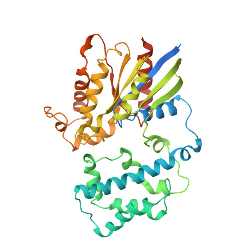
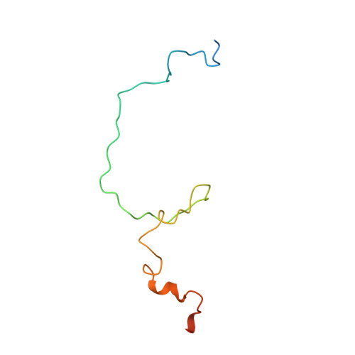
Structure of the Visual Signaling Complex between Transducin and Phosphodiesterase 6.

Gao, Y.Eskici, G.Ramachandran, S.Poitevin, F.Seven, A.B.Panova, O.Skiniotis, G.Cerione, R.A.

(2020) Mol Cell 80: 237

  • DOI: https://doi.org/10.1016/j.molcel.2020.09.013
  • Primary Citation of Related Structures:  
    7JSN

  • PubMed Abstract: 

    Heterotrimeric G proteins communicate signals from activated G protein-coupled receptors to downstream effector proteins. In the phototransduction pathway responsible for vertebrate vision, the G protein-effector complex is composed of the GTP-bound transducin α subunit (Gα T ·GTP) and the cyclic GMP (cGMP) phosphodiesterase 6 (PDE6), which stimulates cGMP hydrolysis, leading to hyperpolarization of the photoreceptor cell. Here we report a cryo-electron microscopy (cryoEM) structure of PDE6 complexed to GTP-bound Gα T . The structure reveals two Gα T ·GTP subunits engaging the PDE6 hetero-tetramer at both the PDE6 catalytic core and the PDEγ subunits, driving extensive rearrangements to relieve all inhibitory constraints on enzyme catalysis. Analysis of the conformational ensemble in the cryoEM data highlights the dynamic nature of the contacts between the two Gα T ·GTP subunits and PDE6 that supports an alternating-site catalytic mechanism.


  • Organizational Affiliation

    Department of Molecular and Cellular Physiology, Stanford University School of Medicine, Stanford, CA 94305, USA; Department of Structural Biology, Stanford University School of Medicine, Stanford, CA 94305, USA.


Macromolecules
Find similar proteins by:  (by identity cutoff)  |  3D Structure
Entity ID: 1
MoleculeChains Sequence LengthOrganismDetailsImage
Rod cGMP-specific 3',5'-cyclic phosphodiesterase subunit alpha859Bos taurusMutation(s): 0 
EC: 3.1.4.35
Membrane Entity: Yes 
UniProt
Find proteins for P11541 (Bos taurus)
Explore P11541 
Go to UniProtKB:  P11541
Entity Groups  
Sequence Clusters30% Identity50% Identity70% Identity90% Identity95% Identity100% Identity
UniProt GroupP11541
Sequence Annotations
Expand
  • Reference Sequence
Find similar proteins by:  (by identity cutoff)  |  3D Structure
Entity ID: 2
MoleculeChains Sequence LengthOrganismDetailsImage
Rod cGMP-specific 3',5'-cyclic phosphodiesterase subunit beta853Bos taurusMutation(s): 0 
EC: 3.1.4.35
Membrane Entity: Yes 
UniProt
Find proteins for P23439 (Bos taurus)
Explore P23439 
Go to UniProtKB:  P23439
Entity Groups  
Sequence Clusters30% Identity50% Identity70% Identity90% Identity95% Identity100% Identity
UniProt GroupP23439
Sequence Annotations
Expand
  • Reference Sequence
Find similar proteins by:  (by identity cutoff)  |  3D Structure
Entity ID: 3
MoleculeChains Sequence LengthOrganismDetailsImage
Guanine nucleotide-binding protein G(t) subunit alpha-1, Guanine nucleotide-binding protein G(i) subunit alpha-1 chimeraC [auth E],
D [auth F]
371Bos taurusMutation(s): 2 
Gene Names: GNAT1
Membrane Entity: Yes 
UniProt
Find proteins for P04695 (Bos taurus)
Explore P04695 
Go to UniProtKB:  P04695
Entity Groups  
Sequence Clusters30% Identity50% Identity70% Identity90% Identity95% Identity100% Identity
UniProt GroupP04695
Sequence Annotations
Expand
  • Reference Sequence
Find similar proteins by:  (by identity cutoff)  |  3D Structure
Entity ID: 4
MoleculeChains Sequence LengthOrganismDetailsImage
Retinal rod rhodopsin-sensitive cGMP 3',5'-cyclic phosphodiesterase subunit gammaE [auth C],
F [auth D]
87Bos taurusMutation(s): 0 
EC: 3.1.4.35
UniProt
Find proteins for P04972 (Bos taurus)
Explore P04972 
Go to UniProtKB:  P04972
Entity Groups  
Sequence Clusters30% Identity50% Identity70% Identity90% Identity95% Identity100% Identity
UniProt GroupP04972
Sequence Annotations
Expand
  • Reference Sequence
Small Molecules
Ligands 5 Unique
IDChains Name / Formula / InChI Key2D Diagram3D Interactions
GTP
Query on GTP

Download Ideal Coordinates CCD File 
O [auth E],
P [auth F]
GUANOSINE-5'-TRIPHOSPHATE
C10 H16 N5 O14 P3
XKMLYUALXHKNFT-UUOKFMHZSA-N
VDN (Subject of Investigation/LOI)
Query on VDN

Download Ideal Coordinates CCD File 
I [auth A],
M [auth B]
2-{2-ETHOXY-5-[(4-ETHYLPIPERAZIN-1-YL)SULFONYL]PHENYL}-5-METHYL-7-PROPYLIMIDAZO[5,1-F][1,2,4]TRIAZIN-4(1H)-ONE
C23 H32 N6 O4 S
SECKRCOLJRRGGV-UHFFFAOYSA-N
35G
Query on 35G

Download Ideal Coordinates CCD File 
J [auth A],
N [auth B]
GUANOSINE-3',5'-MONOPHOSPHATE
C10 H12 N5 O7 P
ZOOGRGPOEVQQDX-UUOKFMHZSA-N
ZN
Query on ZN

Download Ideal Coordinates CCD File 
G [auth A],
K [auth B]
ZINC ION
Zn
PTFCDOFLOPIGGS-UHFFFAOYSA-N
MG
Query on MG

Download Ideal Coordinates CCD File 
H [auth A],
L [auth B]
MAGNESIUM ION
Mg
JLVVSXFLKOJNIY-UHFFFAOYSA-N
Experimental Data & Validation

Experimental Data

  • Method: ELECTRON MICROSCOPY
  • Resolution: 3.20 Å
  • Aggregation State: PARTICLE 
  • Reconstruction Method: SINGLE PARTICLE 

Structure Validation

View Full Validation Report


Entry History & Funding Information

Deposition Data


Funding OrganizationLocationGrant Number
National Institutes of Health/National Institute of General Medical Sciences (NIH/NIGMS)United StatesR35 GM122575
National Institutes of Health/National Cancer Institute (NIH/NCI)United StatesR01 CA201402
National Institutes of Health/National Institute of Neurological Disorders and Stroke (NIH/NINDS)United StatesR01 NS092695

Revision History  (Full details and data files)

  • Version 1.0: 2020-10-21
    Type: Initial release
  • Version 1.1: 2020-10-28
    Changes: Database references
  • Version 2.0: 2021-03-31
    Type: Coordinate replacement
    Reason: Model orientation/position
    Changes: Advisory, Atomic model, Author supporting evidence, Data collection, Database references, Derived calculations, Polymer sequence, Refinement description, Source and taxonomy, Structure summary