7JHG

Cryo-EM structure of ATP-bound fully inactive AMPK in complex with Dorsomorphin (Compound C) and Fab-nanobody


Experimental Data Snapshot

  • Method: ELECTRON MICROSCOPY
  • Resolution: 3.47 Å
  • Aggregation State: PARTICLE 
  • Reconstruction Method: SINGLE PARTICLE 

wwPDB Validation   3D Report Full Report


This is version 1.1 of the entry. See complete history


Literature

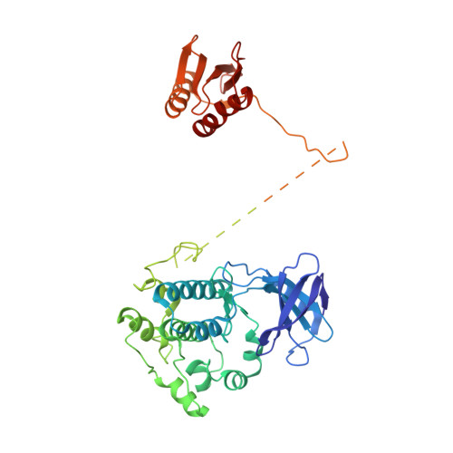
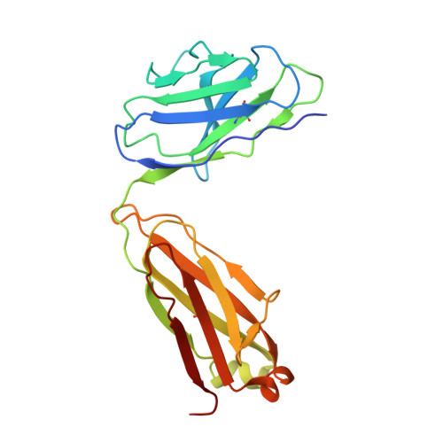
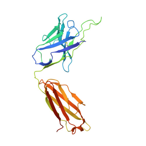
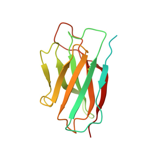
Structure of an AMPK complex in an inactive, ATP-bound state.

Yan, Y.Mukherjee, S.Harikumar, K.G.Strutzenberg, T.S.Zhou, X.E.Suino-Powell, K.Xu, T.H.Sheldon, R.D.Lamp, J.Brunzelle, J.S.Radziwon, K.Ellis, A.Novick, S.J.Vega, I.E.Jones, R.G.Miller, L.J.Xu, H.E.Griffin, P.R.Kossiakoff, A.A.Melcher, K.

(2021) Science 373: 413-419

  • DOI: https://doi.org/10.1126/science.abe7565
  • Primary Citation of Related Structures:  
    7JHG, 7JHH, 7JIJ, 7M74

  • PubMed Abstract: 

    Adenosine monophosphate (AMP)-activated protein kinase (AMPK) regulates metabolism in response to the cellular energy states. Under energy stress, AMP stabilizes the active AMPK conformation, in which the kinase activation loop (AL) is protected from protein phosphatases, thus keeping the AL in its active, phosphorylated state. At low AMP:ATP (adenosine triphosphate) ratios, ATP inhibits AMPK by increasing AL dynamics and accessibility. We developed conformation-specific antibodies to trap ATP-bound AMPK in a fully inactive, dynamic state and determined its structure at 3.5-angstrom resolution using cryo-electron microscopy. A 180° rotation and 100-angstrom displacement of the kinase domain fully exposes the AL. On the basis of the structure and supporting biophysical data, we propose a multistep mechanism explaining how adenine nucleotides and pharmacological agonists modulate AMPK activity by altering AL phosphorylation and accessibility.


  • Organizational Affiliation

    Department of Structural Biology, Van Andel Institute, Grand Rapids, MI 49503, USA.


Macromolecules
Find similar proteins by:  (by identity cutoff)  |  3D Structure
Entity ID: 1
MoleculeChains Sequence LengthOrganismDetailsImage
5'-AMP-activated protein kinase catalytic subunit alpha-1484Homo sapiensMutation(s): 0 
Gene Names: PRKAA1AMPK1
EC: 2.7.11.1 (PDB Primary Data), 2.7.11.27 (PDB Primary Data), 2.7.11.31 (PDB Primary Data), 2.7.11.26 (PDB Primary Data)
UniProt & NIH Common Fund Data Resources
Find proteins for Q13131 (Homo sapiens)
Explore Q13131 
Go to UniProtKB:  Q13131
PHAROS:  Q13131
GTEx:  ENSG00000132356 
Entity Groups  
Sequence Clusters30% Identity50% Identity70% Identity90% Identity95% Identity100% Identity
UniProt GroupQ13131
Sequence Annotations
Expand
  • Reference Sequence
Find similar proteins by:  (by identity cutoff)  |  3D Structure
Entity ID: 2
MoleculeChains Sequence LengthOrganismDetailsImage
5'-AMP-activated protein kinase subunit beta-2198Homo sapiensMutation(s): 0 
Gene Names: PRKAB2
UniProt & NIH Common Fund Data Resources
Find proteins for O43741 (Homo sapiens)
Explore O43741 
Go to UniProtKB:  O43741
PHAROS:  O43741
GTEx:  ENSG00000131791 
Entity Groups  
Sequence Clusters30% Identity50% Identity70% Identity90% Identity95% Identity100% Identity
UniProt GroupO43741
Sequence Annotations
Expand
  • Reference Sequence
Find similar proteins by:  (by identity cutoff)  |  3D Structure
Entity ID: 3
MoleculeChains Sequence LengthOrganismDetailsImage
5'-AMP-activated protein kinase subunit gamma-1C [auth G]306Homo sapiensMutation(s): 0 
Gene Names: PRKAG1
UniProt & NIH Common Fund Data Resources
Find proteins for P54619 (Homo sapiens)
Explore P54619 
Go to UniProtKB:  P54619
PHAROS:  P54619
GTEx:  ENSG00000181929 
Entity Groups  
Sequence Clusters30% Identity50% Identity70% Identity90% Identity95% Identity100% Identity
UniProt GroupP54619
Sequence Annotations
Expand
  • Reference Sequence
Find similar proteins by:  (by identity cutoff)  |  3D Structure
Entity ID: 4
MoleculeChains Sequence LengthOrganismDetailsImage
Maltodextrin-binding proteinD [auth M]373Escherichia coli K-12Mutation(s): 0 
Gene Names: malE
UniProt
Find proteins for P0AEX9 (Escherichia coli (strain K12))
Explore P0AEX9 
Go to UniProtKB:  P0AEX9
Entity Groups  
Sequence Clusters30% Identity50% Identity70% Identity90% Identity95% Identity100% Identity
UniProt GroupP0AEX9
Sequence Annotations
Expand
  • Reference Sequence
Find similar proteins by:  (by identity cutoff)  |  3D Structure
Entity ID: 5
MoleculeChains Sequence LengthOrganismDetailsImage
Fab light chainE [auth L]215synthetic constructMutation(s): 0 
Entity Groups  
Sequence Clusters30% Identity50% Identity70% Identity90% Identity95% Identity100% Identity
Sequence Annotations
Expand
  • Reference Sequence
Find similar proteins by:  (by identity cutoff)  |  3D Structure
Entity ID: 6
MoleculeChains Sequence LengthOrganismDetailsImage
Fab heavy chainF [auth H]237synthetic constructMutation(s): 0 
Entity Groups  
Sequence Clusters30% Identity50% Identity70% Identity90% Identity95% Identity100% Identity
Sequence Annotations
Expand
  • Reference Sequence
Find similar proteins by:  (by identity cutoff)  |  3D Structure
Entity ID: 7
MoleculeChains Sequence LengthOrganismDetailsImage
NanobodyG [auth N]159synthetic constructMutation(s): 0 
Entity Groups  
Sequence Clusters30% Identity50% Identity70% Identity90% Identity95% Identity100% Identity
Sequence Annotations
Expand
  • Reference Sequence
Oligosaccharides

Help

Entity ID: 8
MoleculeChains Length2D Diagram Glycosylation3D Interactions
alpha-D-glucopyranose-(1-4)-alpha-D-glucopyranoseH [auth D]2N/A
Glycosylation Resources
GlyTouCan:  G07411ON
GlyCosmos:  G07411ON
Small Molecules
Ligands 4 Unique
IDChains Name / Formula / InChI Key2D Diagram3D Interactions
ATP
Query on ATP

Download Ideal Coordinates CCD File 
J [auth G]ADENOSINE-5'-TRIPHOSPHATE
C10 H16 N5 O13 P3
ZKHQWZAMYRWXGA-KQYNXXCUSA-N
ADP
Query on ADP

Download Ideal Coordinates CCD File 
K [auth G]ADENOSINE-5'-DIPHOSPHATE
C10 H15 N5 O10 P2
XTWYTFMLZFPYCI-KQYNXXCUSA-N
TAK (Subject of Investigation/LOI)
Query on TAK

Download Ideal Coordinates CCD File 
I [auth A]6-[4-(2-piperidin-1-ylethoxy)phenyl]-3-pyridin-4-ylpyrazolo[1,5-a]pyrimidine
C24 H25 N5 O
XHBVYDAKJHETMP-UHFFFAOYSA-N
AMP
Query on AMP

Download Ideal Coordinates CCD File 
L [auth G]ADENOSINE MONOPHOSPHATE
C10 H14 N5 O7 P
UDMBCSSLTHHNCD-KQYNXXCUSA-N
Biologically Interesting Molecules (External Reference) 1 Unique
Experimental Data & Validation

Experimental Data

  • Method: ELECTRON MICROSCOPY
  • Resolution: 3.47 Å
  • Aggregation State: PARTICLE 
  • Reconstruction Method: SINGLE PARTICLE 
EM Software:
TaskSoftware PackageVersion
MODEL REFINEMENTPHENIX

Structure Validation

View Full Validation Report


Entry History & Funding Information

Deposition Data


Funding OrganizationLocationGrant Number
National Institutes of Health/National Institute of General Medical Sciences (NIH/NIGMS)United StatesR01 GM117372
National Institutes of Health/National Institute of General Medical Sciences (NIH/NIGMS)United StatesR01 GM129436

Revision History  (Full details and data files)

  • Version 1.0: 2021-07-21
    Type: Initial release
  • Version 1.1: 2021-12-15
    Changes: Database references