7F3G

Crystal structure of DCLK1 kinase domain in complex with ruxolitinib


Experimental Data Snapshot

  • Method: X-RAY DIFFRACTION
  • Resolution: 2.10 Å
  • R-Value Free: 0.253 
  • R-Value Work: 0.210 

wwPDB Validation   3D Report Full Report


Ligand Structure Quality Assessment 


This is version 1.2 of the entry. See complete history


Literature

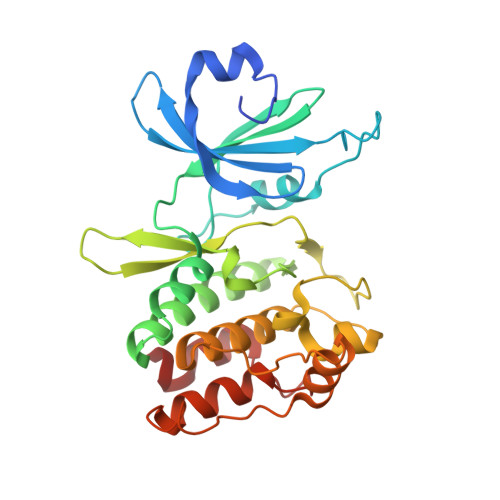
Structural Basis of Inhibition of DCLK1 by Ruxolitinib.

Jang, D.M.Lim, H.J.Hahn, H.Lee, Y.Kim, H.K.Kim, H.S.

(2021) Int J Mol Sci 22

  • DOI: https://doi.org/10.3390/ijms22168488
  • Primary Citation of Related Structures:  
    7F3G

  • PubMed Abstract: 

    Given the functional attributes of Doublecortin-like kinase 1 (DCLK1) in tumor growth, invasion, metastasis, cell motility, and tumor stemness, it is emerging as a therapeutic target in gastrointestinal cancers. Although a series of specific or nonspecific ATP-competitive inhibitors were identified against DCLK1, different types of scaffolds that can be utilized for the development of highly selective inhibitors or structural understanding of binding specificities of the compounds remain limited. Here, we present our work to repurpose a Janus kinase 1 inhibitor, ruxolitinib as a DCLK1 inhibitor, showing micromolar binding affinity and inhibitory activity. Furthermore, to gain an insight into its interaction mode with DCLK1, a crystal structure of the ruxolitinib-complexed DCLK1 has been determined and analyzed. Ruxolitinib as a nonspecific DCLK1 inhibitor characterized in this work is anticipated to provide a starting point for the structure-guided discovery of selective DCLK1 inhibitors.


  • Organizational Affiliation

    Research Institute, National Cancer Center, Goyang 10408, Gyeonggi, Korea.


Macromolecules
Find similar proteins by:  (by identity cutoff)  |  3D Structure
Entity ID: 1
MoleculeChains Sequence LengthOrganismDetailsImage
Isoform 4 of Serine/threonine-protein kinase DCLK1
A, B
282Homo sapiensMutation(s): 0 
Gene Names: DCLK1DCAMKL1DCDC3AKIAA0369
EC: 2.7.11.1
UniProt & NIH Common Fund Data Resources
Find proteins for O15075 (Homo sapiens)
Explore O15075 
Go to UniProtKB:  O15075
PHAROS:  O15075
GTEx:  ENSG00000133083 
Entity Groups  
Sequence Clusters30% Identity50% Identity70% Identity90% Identity95% Identity100% Identity
UniProt GroupO15075
Sequence Annotations
Expand
  • Reference Sequence
Small Molecules
Ligands 1 Unique
IDChains Name / Formula / InChI Key2D Diagram3D Interactions
RXT (Subject of Investigation/LOI)
Query on RXT

Download Ideal Coordinates CCD File 
C [auth A],
D [auth B]
(3R)-3-cyclopentyl-3-[4-(7H-pyrrolo[2,3-d]pyrimidin-4-yl)-1H-pyrazol-1-yl]propanenitrile
C17 H18 N6
HFNKQEVNSGCOJV-OAHLLOKOSA-N
Experimental Data & Validation

Experimental Data

  • Method: X-RAY DIFFRACTION
  • Resolution: 2.10 Å
  • R-Value Free: 0.253 
  • R-Value Work: 0.210 
  • Space Group: P 1 21 1
Unit Cell:
Length ( Å )Angle ( ˚ )
a = 57.248α = 90
b = 80.118β = 90.662
c = 59.338γ = 90
Software Package:
Software NamePurpose
REFMACrefinement
XDSdata reduction
XDSdata scaling
MOLREPphasing

Structure Validation

View Full Validation Report


Ligand Structure Quality Assessment 


Entry History & Funding Information

Deposition Data


Funding OrganizationLocationGrant Number
National Research Foundation (NRF, Korea)Korea, Republic OfNRF-2020R1C1C1009512

Revision History  (Full details and data files)

  • Version 1.0: 2021-08-18
    Type: Initial release
  • Version 1.1: 2022-04-27
    Changes: Database references
  • Version 1.2: 2023-11-29
    Changes: Data collection, Derived calculations, Refinement description