Structural basis for substrate specificity of the peroxisomal acyl-CoA hydrolase MpaH' involved in mycophenolic acid biosynthesis.
You, C., Li, F., Zhang, X., Ma, L., Zhang, Y.Z., Zhang, W., Li, S.(2021) FEBS J 288: 5768-5780
- PubMed: 33843134
- DOI: https://doi.org/10.1111/febs.15874
- Primary Citation of Related Structures:
7DBI, 7DBL - PubMed Abstract:
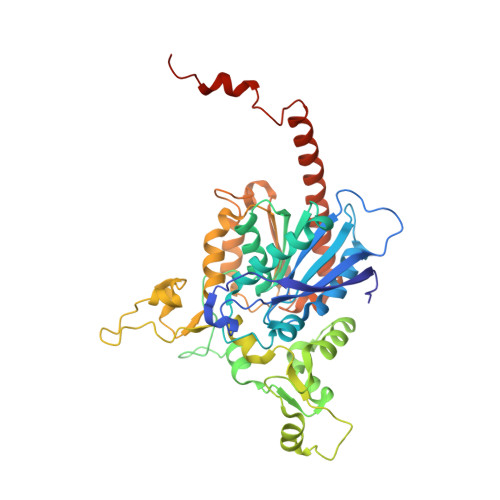
Mycophenolic acid (MPA) is a fungal natural product and first-line immunosuppressive drug for organ transplantations and autoimmune diseases. In the compartmentalized biosynthesis of MPA, the acyl-coenzyme A (CoA) hydrolase MpaH' located in peroxisomes catalyzes the highly specific hydrolysis of MPA-CoA to produce the final product MPA. The strict substrate specificity of MpaH' not only averts undesired hydrolysis of various cellular acyl-CoAs, but also prevents MPA-CoA from further peroxisomal β-oxidation catabolism. To elucidate the structural basis for this important property, in this study, we solve the crystal structures of the substrate-free form of MpaH' and the MpaH' S139A mutant in complex with the product MPA. The MpaH' structure reveals a canonical α/β-hydrolase fold with an unusually large cap domain and a rare location of the acidic residue D163 of catalytic triad after strand β6. MpaH' also forms an atypical dimer with the unique C-terminal helices α13 and α14 arming the cap domain of the other protomer and indirectly participating in the substrate binding. With these characteristics, we propose that MpaH' and its homologs form a new subfamily of α/β hydrolase fold protein. The crystal structure of MpaH' S139A /MPA complex and the modeled structure of MpaH'/MPA-CoA, together with the structure-guided mutagenesis analysis and isothermal titration calorimetry (ITC) measurements, provide important mechanistic insights into the high substrate specificity of MpaH'.
Organizational Affiliation:
State Key Laboratory of Microbial Technology, Shandong University, Qingdao, China.