7CEP

Crystal structure of L-cycloserine-bound form of cysteine desulfurase SufS from Bacillus subtilis


Experimental Data Snapshot

  • Method: X-RAY DIFFRACTION
  • Resolution: 2.05 Å
  • R-Value Free: 0.186 
  • R-Value Work: 0.166 
  • R-Value Observed: 0.167 

wwPDB Validation   3D Report Full Report


Ligand Structure Quality Assessment 


This is version 1.2 of the entry. See complete history


Literature

Cycloserine enantiomers inhibit PLP-dependent cysteine desulfurase SufS via distinct mechanisms.

Nakamura, R.Ogawa, S.Takahashi, Y.Fujishiro, T.

(2022) FEBS J 

  • DOI: https://doi.org/10.1111/febs.16455
  • Primary Citation of Related Structures:  
    6KFY, 7CEO, 7CEP, 7CEQ, 7CER, 7CES, 7CET, 7CEU, 7E6A, 7E6B, 7E6C, 7E6D, 7E6E, 7E6F

  • PubMed Abstract: 

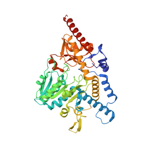
    The cysteine desulfurase SufS is a pyridoxal-5'-phosphate-dependent enzyme and is essential for the SUF system, which participates in iron-sulfur cluster biosynthesis. Inhibition of SufS in the SUF system by D-cycloserine (DCS) in Plasmodium falciparum apicoplast has recently been reported, indicating that SufS could be a target for malaria therapeutics. However, the mechanistic details underlying the inhibition of SufS by DCS have not yet been clarified. Moreover, inhibition of SufS by the other enantiomer, L-cycloserine (LCS), has not been investigated. Herein, we investigated the structure-based inhibition mechanisms of SufS by DCS and LCS using Bacillus subtilis SufS, whose catalytic mechanism has been well characterized in comparison to that of the P. falciparum SufS. Surprisingly, DCS- and LCS-mediated inhibitions of SufS occur via distinct mechanisms resulting in pyridoxamine-5'-phosphate (PMP) in DCS-mediated inhibition and PMP-3-hydroxyisoxazole adduct (PMP-isoxazole) in LCS-mediated inhibition. Biochemical and structural evaluation of SufS variants identified conserved His and Arg residues at the active site as the key determinants of the distinct inhibition mechanisms. The importance of structural elements involved in DCS and LCS-mediated inhibitions of SufS provides valuable insights for the structure-based design of new drugs targeting SufS. DATABASE: Structural data are available in PDB database under the accession numbers 6KFY, 7CEO, 7CEP, 7CEQ, 7CER, 7CES, 7CET, 7CEU, 7E6A, 7E6B, 7E6C, 7E6D, 7E6E, and 7E6F.


  • Organizational Affiliation

    Department of Biochemistry and Molecular Biology, Graduate School of Science and Engineering, Saitama University, Japan.


Macromolecules
Find similar proteins by:  (by identity cutoff)  |  3D Structure
Entity ID: 1
MoleculeChains Sequence LengthOrganismDetailsImage
Cysteine desulfurase SufS419Bacillus subtilis subsp. subtilis str. 168Mutation(s): 0 
Gene Names: sufScsdyurWBSU32690
EC: 2.8.1.7
UniProt
Find proteins for O32164 (Bacillus subtilis (strain 168))
Explore O32164 
Go to UniProtKB:  O32164
Entity Groups  
Sequence Clusters30% Identity50% Identity70% Identity90% Identity95% Identity100% Identity
UniProt GroupO32164
Sequence Annotations
Expand
  • Reference Sequence
Small Molecules
Ligands 3 Unique
IDChains Name / Formula / InChI Key2D Diagram3D Interactions
7TS (Subject of Investigation/LOI)
Query on 7TS

Download Ideal Coordinates CCD File 
H [auth A](5-hydroxy-6-methyl-4-{[(3-oxo-2,3-dihydro-1,2-oxazol-4-yl)amino]methyl}pyridin-3-yl)methyl dihydrogen phosphate
C11 H14 N3 O7 P
PXWFNGNWQUPGPJ-UHFFFAOYSA-N
PGE
Query on PGE

Download Ideal Coordinates CCD File 
I [auth A]TRIETHYLENE GLYCOL
C6 H14 O4
ZIBGPFATKBEMQZ-UHFFFAOYSA-N
PEG
Query on PEG

Download Ideal Coordinates CCD File 
B [auth A]
C [auth A]
D [auth A]
E [auth A]
F [auth A]
B [auth A],
C [auth A],
D [auth A],
E [auth A],
F [auth A],
G [auth A]
DI(HYDROXYETHYL)ETHER
C4 H10 O3
MTHSVFCYNBDYFN-UHFFFAOYSA-N
Experimental Data & Validation

Experimental Data

  • Method: X-RAY DIFFRACTION
  • Resolution: 2.05 Å
  • R-Value Free: 0.186 
  • R-Value Work: 0.166 
  • R-Value Observed: 0.167 
  • Space Group: P 31 2 1
Unit Cell:
Length ( Å )Angle ( ˚ )
a = 92.8α = 90
b = 92.8β = 90
c = 128.68γ = 120
Software Package:
Software NamePurpose
XSCALEdata scaling
PHENIXrefinement
PDB_EXTRACTdata extraction
XDSdata reduction
MOLREPphasing

Structure Validation

View Full Validation Report


Ligand Structure Quality Assessment 


Entry History & Funding Information

Deposition Data


Funding OrganizationLocationGrant Number
Japan Society for the Promotion of Science (JSPS)Japan17K14510

Revision History  (Full details and data files)

  • Version 1.0: 2021-06-30
    Type: Initial release
  • Version 1.1: 2022-04-27
    Changes: Database references
  • Version 1.2: 2023-11-29
    Changes: Data collection, Refinement description