7BVH

Crystal structure of arabinosyltransferase EmbC2-AcpM2 complex from Mycobacterium smegmatis complexed with di-arabinose


Experimental Data Snapshot

  • Method: X-RAY DIFFRACTION
  • Resolution: 3.30 Å
  • R-Value Free: 0.265 
  • R-Value Work: 0.232 
  • R-Value Observed: 0.234 

wwPDB Validation   3D Report Full Report


Ligand Structure Quality Assessment 


This is version 2.0 of the entry. See complete history


Literature

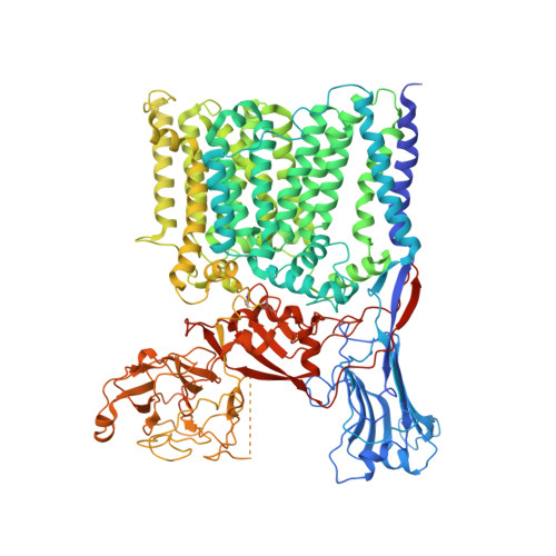
Structures of cell wall arabinosyltransferases with the anti-tuberculosis drug ethambutol.

Zhang, L.Zhao, Y.Gao, Y.Wu, L.Gao, R.Zhang, Q.Wang, Y.Wu, C.Wu, F.Gurcha, S.S.Veerapen, N.Batt, S.M.Zhao, W.Qin, L.Yang, X.Wang, M.Zhu, Y.Zhang, B.Bi, L.Zhang, X.Yang, H.Guddat, L.W.Xu, W.Wang, Q.Li, J.Besra, G.S.Rao, Z.

(2020) Science 368: 1211-1219

  • DOI: https://doi.org/10.1126/science.aba9102
  • Primary Citation of Related Structures:  
    7BVC, 7BVE, 7BVF, 7BVG, 7BVH

  • PubMed Abstract: 

    The arabinosyltransferases EmbA, EmbB, and EmbC are involved in Mycobacterium tuberculosis cell wall synthesis and are recognized as targets for the anti-tuberculosis drug ethambutol. In this study, we determined cryo-electron microscopy and x-ray crystal structures of mycobacterial EmbA-EmbB and EmbC-EmbC complexes in the presence of their glycosyl donor and acceptor substrates and with ethambutol. These structures show how the donor and acceptor substrates bind in the active site and how ethambutol inhibits arabinosyltransferases by binding to the same site as both substrates in EmbB and EmbC. Most drug-resistant mutations are located near the ethambutol binding site. Collectively, our work provides a structural basis for understanding the biochemical function and inhibition of arabinosyltransferases and the development of new anti-tuberculosis agents.


  • Organizational Affiliation

    Shanghai Institute for Advanced Immunochemical Studies, iHuman Institute, School of Life Science and Technology, ShanghaiTech University, Shanghai 201210, China.


Macromolecules
Find similar proteins by:  (by identity cutoff)  |  3D Structure
Entity ID: 1
MoleculeChains Sequence LengthOrganismDetailsImage
Integral membrane indolylacetylinositol arabinosyltransferase EmbC
A, B
1,113Mycolicibacterium smegmatis MC2 155Mutation(s): 0 
Gene Names: embCMSMEI_6219
EC: 2.4.2.34
Membrane Entity: Yes 
UniProt
Find proteins for A0R612 (Mycolicibacterium smegmatis (strain ATCC 700084 / mc(2)155))
Explore A0R612 
Go to UniProtKB:  A0R612
Entity Groups  
Sequence Clusters30% Identity50% Identity70% Identity90% Identity95% Identity100% Identity
UniProt GroupA0R612
Sequence Annotations
Expand
  • Reference Sequence
Find similar proteins by:  (by identity cutoff)  |  3D Structure
Entity ID: 2
MoleculeChains Sequence LengthOrganismDetailsImage
Meromycolate extension acyl carrier protein
C, D
99Mycolicibacterium smegmatis MC2 155Mutation(s): 0 
UniProt
Find proteins for A0R0B3 (Mycolicibacterium smegmatis (strain ATCC 700084 / mc(2)155))
Explore A0R0B3 
Go to UniProtKB:  A0R0B3
Entity Groups  
Sequence Clusters30% Identity50% Identity70% Identity90% Identity95% Identity100% Identity
UniProt GroupA0R0B3
Sequence Annotations
Expand
  • Reference Sequence
Oligosaccharides

Help

Entity ID: 3
MoleculeChains Length2D Diagram Glycosylation3D Interactions
alpha-D-arabinofuranose-(1-5)-alpha-D-arabinofuranose
E, J
2N/A
Glycosylation Resources
GlyTouCan:  G34232WB
GlyCosmos:  G34232WB
Entity ID: 4
MoleculeChains Length2D Diagram Glycosylation3D Interactions
alpha-D-glucopyranose-(1-4)-beta-D-glucopyranose
F, G, H, I, K
F, G, H, I, K, L
2N/A
Glycosylation Resources
GlyTouCan:  G66120GK
GlyCosmos:  G66120GK
Small Molecules
Ligands 3 Unique
IDChains Name / Formula / InChI Key2D Diagram3D Interactions
BXY
Query on BXY

Download Ideal Coordinates CCD File 
O [auth A],
P [auth A],
S [auth B],
T [auth B]
alpha-D-arabinofuranose
C5 H10 O5
HMFHBZSHGGEWLO-MBMOQRBOSA-N
PO4 (Subject of Investigation/LOI)
Query on PO4

Download Ideal Coordinates CCD File 
N [auth A],
R [auth B]
PHOSPHATE ION
O4 P
NBIIXXVUZAFLBC-UHFFFAOYSA-K
CA
Query on CA

Download Ideal Coordinates CCD File 
M [auth A],
Q [auth B]
CALCIUM ION
Ca
BHPQYMZQTOCNFJ-UHFFFAOYSA-N
Experimental Data & Validation

Experimental Data

  • Method: X-RAY DIFFRACTION
  • Resolution: 3.30 Å
  • R-Value Free: 0.265 
  • R-Value Work: 0.232 
  • R-Value Observed: 0.234 
  • Space Group: P 21 21 21
Unit Cell:
Length ( Å )Angle ( ˚ )
a = 121.08α = 90
b = 176.33β = 90
c = 207.77γ = 90
Software Package:
Software NamePurpose
BUSTERrefinement
SCALAdata scaling
PDB_EXTRACTdata extraction
XDSdata reduction
PHENIXphasing

Structure Validation

View Full Validation Report


Ligand Structure Quality Assessment 


Entry History & Funding Information

Deposition Data


Funding OrganizationLocationGrant Number
Ministry of Science and Technology (MoST, China)China2017YFC0840300
National Science Foundation (NSF, China)China81520108019
Chinese Academy of SciencesChinaXDB29020000
Medical Research Council (MRC, United Kingdom)United KingdomMR/S000542/1

Revision History  (Full details and data files)

  • Version 1.0: 2020-04-29
    Type: Initial release
  • Version 1.1: 2020-05-13
    Changes: Database references
  • Version 1.2: 2020-07-01
    Changes: Database references
  • Version 2.0: 2020-07-29
    Type: Remediation
    Reason: Carbohydrate remediation
    Changes: Advisory, Atomic model, Data collection, Derived calculations, Structure summary