


Crystal structure of a tRNA (guanine-N1)-methyltransferase from Acinetobacter baumannii
Edwards, T.E., Abendroth, J., Horanyi, P.S., Lorimer, D.D., Seattle Structural Genomics Center for Infectious DiseaseTo be published.
Experimental Data Snapshot
wwPDB Validation 3D Report Full Report

Entity ID: 1 | |||||
|---|---|---|---|---|---|
| Molecule | Chains | Sequence Length | Organism | Details | Image |
| tRNA (guanine-N(1)-)-methyltransferase | 254 | Acinetobacter baumannii | Mutation(s): 0 Gene Names: trmD, ABC003_01337, ABUW_0322, EA686_14030, FDO31_01140, FJU79_00125, GNY86_14875, HYI42_11650 EC: 2.1.1.228 | 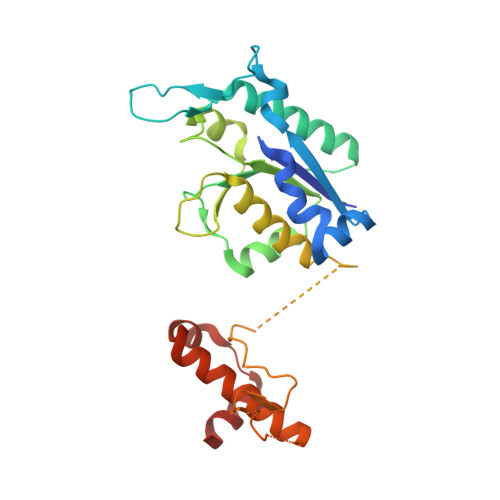 | |
UniProt | |||||
Find proteins for B0V8J1 (Acinetobacter baumannii (strain AYE)) Explore B0V8J1 Go to UniProtKB: B0V8J1 | |||||
Entity Groups | |||||
| Sequence Clusters | 30% Identity50% Identity70% Identity90% Identity95% Identity100% Identity | ||||
| UniProt Group | B0V8J1 | ||||
Sequence AnnotationsExpand | |||||
| |||||
| Length ( Å ) | Angle ( ˚ ) |
|---|---|
| a = 66.25 | α = 90 |
| b = 66.25 | β = 90 |
| c = 133.01 | γ = 90 |
| Software Name | Purpose |
|---|---|
| XDS | data reduction |
| XSCALE | data scaling |
| MOLREP | phasing |
| PHENIX | refinement |
| PDB_EXTRACT | data extraction |
RCSB PDB (citation) is hosted by
RCSB PDB is a member of the

