Structural and biochemical characterization of the class II fructose-1,6-bisphosphatase from Francisella tularensis.
Selezneva, A.I., Gutka, H.J., Wolf, N.M., Qurratulain, F., Movahedzadeh, F., Abad-Zapatero, C.(2020) Acta Crystallogr F Struct Biol Commun 76: 524-535
- PubMed: 33135671
- DOI: https://doi.org/10.1107/S2053230X20013370
- Primary Citation of Related Structures:
7JS3 - PubMed Abstract:
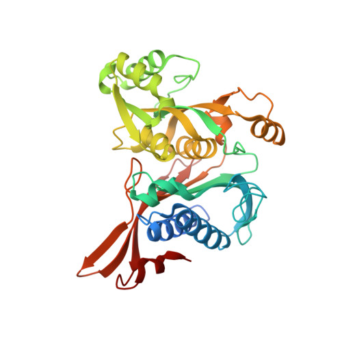
The crystal structure of the class II fructose-1,6-bisphosphatase (FBPaseII) from the important pathogen Francisella tularensis is presented at 2.4 Å resolution. Its structural and functional relationships to the closely related phosphatases from Mycobacterium tuberculosis (MtFBPaseII) and Escherichia coli (EcFBPaseII) and to the dual phosphatase from Synechocystis strain 6803 are discussed. FBPaseII from F. tularensis (FtFBPaseII) was crystallized in a monoclinic crystal form (space group P2 1 , unit-cell parameters a = 76.30, b = 100.17, c = 92.02 Å, β = 90.003°) with four chains in the asymmetric unit. Chain A had two coordinated Mg 2+ ions in its active center, which is distinct from previous findings, and is presumably deactivated by their presence. The structure revealed an approximate 222 (D 2 ) symmetry homotetramer analogous to that previously described for MtFBPaseII, which is formed by a crystallographic dyad and which differs from the exact tetramer found in EcFBPaseII at a 222 symmetry site in the crystal. Instead, the approximate homotetramer is very similar to that found in the dual phosphatase from Synechocystis, even though no allosteric effector was found in FtFBPase. The amino-acid sequence and folding of the active site of FtFBPaseII result in structural characteristics that are more similar to those of the previously published EcFBPaseII than to those of MtFBPaseII. The kinetic parameters of native FtFBPaseII were found to be in agreement with published studies. Kinetic analyses of the Thr89Ser and Thr89Ala mutations in the active site of the enzyme are consistent with the previously proposed mechanism for other class II bisphosphatases. The Thr89Ala variant enzyme was inactive but the Thr89Ser variant was partially active, with an approximately fourfold lower K m and V max than the native enzyme. The structural and functional insights derived from the structure of FtFBPaseII will provide valuable information for the design of specific inhibitors.
Organizational Affiliation:
Institute for Tuberculosis Research, University of Illinois at Chicago, Chicago, Illinois, USA.