7EXU

GH127 beta-L-arabinofuranosidase HypBA1 E322Q mutant complexed with p-nitrophenyl beta-L-arabinofuranoside


Experimental Data Snapshot

  • Method: X-RAY DIFFRACTION
  • Resolution: 2.30 Å
  • R-Value Free: 0.290 
  • R-Value Work: 0.252 
  • R-Value Observed: 0.254 

wwPDB Validation   3D Report Full Report


Ligand Structure Quality Assessment 


This is version 1.3 of the entry. See complete history


Literature

Substrate complex structure, active site labeling and catalytic role of the zinc ion in cysteine glycosidase.

Maruyama, S.Sawano, K.Amaki, S.Suzuki, T.Narita, S.Kimura, K.Arakawa, T.Yamada, C.Ito, Y.Dohmae, N.Fujita, K.Ishiwata, A.Fushinobu, S.

(2022) Glycobiology 32: 171-180

  • DOI: https://doi.org/10.1093/glycob/cwab103
  • Primary Citation of Related Structures:  
    7EXU, 7EXV, 7EXW

  • PubMed Abstract: 

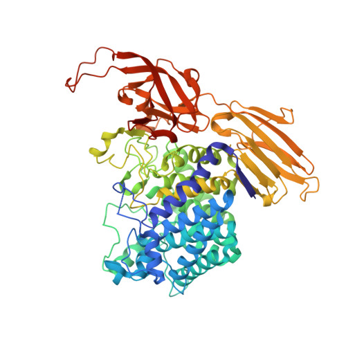
    β-l-Arabinofuranosidase HypBA1 from Bifidobacterium longum belongs to the glycoside hydrolase family 127. At the active site of HypBA1, a cysteine residue (Cys417) coordinates with a Zn2+ atom and functions as the catalytic nucleophile for the anomer-retaining hydrolytic reaction. In this study, the role of Zn2+ ion and cysteine in catalysis as well as the substrate-bound structure were studied based on biochemical and crystallographic approaches. The enzymatic activity of HypBA1 decreased after dialysis in the presence of EDTA and guanidine hydrochloride and was then recovered by the addition of Zn2+. The Michaelis complex structure was determined using a crystal of a mutant at the acid/base catalyst residue (E322Q) soaked in a solution containing the substrate p-nitrophenyl-β-l-arabinofuranoside. To investigate the covalent thioglycosyl enzyme intermediate structure, synthetic inhibitors of l-arabinofuranosyl haloacetamide derivatives with different anomer configurations were used to target the nucleophilic cysteine. In the crystal structure of HypBA1, β-configured l-arabinofuranosylamide formed a covalent link with Cys417, whereas α-configured l-arabinofuranosylamide was linked to a noncatalytic residue Cys415. Mass spectrometric analysis indicated that Cys415 was also reactive with the probe molecule. With the β-configured inhibitor, the arabinofuranoside moiety was correctly positioned at the subsite and the active site integrity was retained to successfully mimic the covalent intermediate state.


  • Organizational Affiliation

    Department of Biotechnology, The University of Tokyo, 1-1-1 Yayoi, Bunkyo-ku, Tokyo 113-8657, Japan.


Macromolecules
Find similar proteins by:  (by identity cutoff)  |  3D Structure
Entity ID: 1
MoleculeChains Sequence LengthOrganismDetailsImage
Non-reducing end beta-L-arabinofuranosidase669Bifidobacterium longum subsp. longum JCM 1217Mutation(s): 1 
Gene Names: hypBA1BLLJ_0211
EC: 3.2.1.185
UniProt
Find proteins for E8MGH8 (Bifidobacterium longum subsp. longum (strain ATCC 15707 / DSM 20219 / JCM 1217 / NCTC 11818 / E194b))
Explore E8MGH8 
Go to UniProtKB:  E8MGH8
Entity Groups  
Sequence Clusters30% Identity50% Identity70% Identity90% Identity95% Identity100% Identity
UniProt GroupE8MGH8
Sequence Annotations
Expand
  • Reference Sequence
Small Molecules
Ligands 2 Unique
IDChains Name / Formula / InChI Key2D Diagram3D Interactions
07Y (Subject of Investigation/LOI)
Query on 07Y

Download Ideal Coordinates CCD File 
C [auth A](2S,3R,4R,5R)-2-(hydroxymethyl)-5-(4-nitrophenoxy)oxolane-3,4-diol
C11 H13 N O7
DUYYBTBDYZXISX-MMWGEVLESA-N
ZN (Subject of Investigation/LOI)
Query on ZN

Download Ideal Coordinates CCD File 
B [auth A]ZINC ION
Zn
PTFCDOFLOPIGGS-UHFFFAOYSA-N
Experimental Data & Validation

Experimental Data

  • Method: X-RAY DIFFRACTION
  • Resolution: 2.30 Å
  • R-Value Free: 0.290 
  • R-Value Work: 0.252 
  • R-Value Observed: 0.254 
  • Space Group: P 32 2 1
Unit Cell:
Length ( Å )Angle ( ˚ )
a = 78.182α = 90
b = 78.182β = 90
c = 253.502γ = 120
Software Package:
Software NamePurpose
REFMACrefinement
PDB_EXTRACTdata extraction
XDSdata reduction
Aimlessdata scaling

Structure Validation

View Full Validation Report


Ligand Structure Quality Assessment 


Entry History & Funding Information

Deposition Data


Funding OrganizationLocationGrant Number
Japan Society for the Promotion of Science (JSPS)Japan15H02443
Japan Society for the Promotion of Science (JSPS)Japan26660083
Japan Society for the Promotion of Science (JSPS)Japan19H00929

Revision History  (Full details and data files)

  • Version 1.0: 2021-11-17
    Type: Initial release
  • Version 1.1: 2022-03-30
    Changes: Database references
  • Version 1.2: 2023-11-29
    Changes: Data collection, Refinement description
  • Version 1.3: 2024-02-21
    Changes: Data collection