


7CWD
Crystal structure of beta-galactosidase II from Bacillus circulans in complex with beta-D-galactopyranosyl disaccharide
- PDB DOI: https://doi.org/10.2210/pdb7CWD/pdb
- Classification: HYDROLASE
- Organism(s): Niallia circulans
- Expression System: Escherichia coli
- Mutation(s): No
- Deposited: 2020-08-27 Released: 2020-12-09
Experimental Data Snapshot
- Method: X-RAY DIFFRACTION
- Resolution: 2.00 Å
- R-Value Free: 0.218
- R-Value Work: 0.189

This is version 1.1 of the entry. See complete history.

Macromolecules
Find similar proteins by:
(by identity cutoff) | 3D Structure
Entity ID: 1 | |||||
|---|---|---|---|---|---|
| Molecule | Chains | Sequence Length | Organism | Details | Image |
| beta-glalactosidase | 815 | Niallia circulans | Mutation(s): 0 EC: 3.2.1.23 | 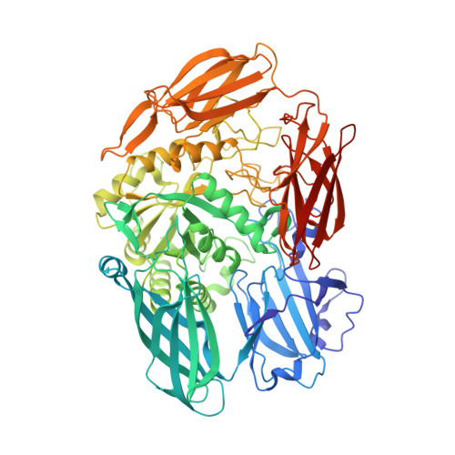 | |
UniProt | |||||
Find proteins for A0A6M5K904 (Niallia circulans) Explore A0A6M5K904 Go to UniProtKB: A0A6M5K904 | |||||
Entity Groups | |||||
| Sequence Clusters | 30% Identity50% Identity70% Identity90% Identity95% Identity100% Identity | ||||
| UniProt Group | A0A6M5K904 | ||||
Sequence AnnotationsExpand | |||||
| |||||
Small Molecules
| Ligands 2 Unique | |||||
|---|---|---|---|---|---|
| ID | Chains | Name / Formula / InChI Key | 2D Diagram | 3D Interactions | |
| GAL (Subject of Investigation/LOI) Query on GAL | C [auth A], D [auth A] | beta-D-galactopyranose C6 H12 O6 WQZGKKKJIJFFOK-FPRJBGLDSA-N |  | ||
| GLC (Subject of Investigation/LOI) Query on GLC | B [auth A] | alpha-D-glucopyranose C6 H12 O6 WQZGKKKJIJFFOK-DVKNGEFBSA-N |  | ||
Experimental Data & Validation
Experimental Data
- Method: X-RAY DIFFRACTION
- Resolution: 2.00 Å
- R-Value Free: 0.218
- R-Value Work: 0.189
- Space Group: P 32 2 1
Unit Cell:
| Length ( Å ) | Angle ( ˚ ) |
|---|---|
| a = 159.499 | α = 90 |
| b = 159.499 | β = 90 |
| c = 96.326 | γ = 120 |
| Software Name | Purpose |
|---|---|
| REFMAC | refinement |
| HKL-2000 | data scaling |
| PDB_EXTRACT | data extraction |
| HKL-2000 | data collection |
| MOLREP | phasing |
| Coot | model building |
| HKL-2000 | data reduction |
Entry History
Revision History (Full details and data files)
- Version 1.0: 2020-12-09
Type: Initial release - Version 1.1: 2023-11-29
Changes: Data collection, Database references, Refinement description
