6ZPV

Structure of Unliganded MgGH51 a-L-Arabinofuranosidase Crystal Type 3


Experimental Data Snapshot

  • Method: X-RAY DIFFRACTION
  • Resolution: 1.20 Å
  • R-Value Free: 0.144 
  • R-Value Work: 0.120 

wwPDB Validation   3D Report Full Report


This is version 1.2 of the entry. See complete history


Literature

Structure of a GH51 alpha-L-arabinofuranosidase from Meripilus giganteus: conserved substrate recognition from bacteria to fungi.

McGregor, N.G.S.Turkenburg, J.P.Morkeberg Krogh, K.B.R.Nielsen, J.E.Artola, M.Stubbs, K.A.Overkleeft, H.S.Davies, G.J.

(2020) Acta Crystallogr D Struct Biol 76: 1124-1133

  • DOI: https://doi.org/10.1107/S205979832001253X
  • Primary Citation of Related Structures:  
    6ZPS, 6ZPV, 6ZPW, 6ZPX, 6ZPY, 6ZPZ, 6ZQ0, 6ZQ1

  • PubMed Abstract: 

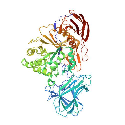
    α-L-Arabinofuranosidases from glycoside hydrolase family 51 use a stereochemically retaining hydrolytic mechanism to liberate nonreducing terminal α-L-arabinofuranose residues from plant polysaccharides such as arabinoxylan and arabinan. To date, more than ten fungal GH51 α-L-arabinofuranosidases have been functionally characterized, yet no structure of a fungal GH51 enzyme has been solved. In contrast, seven bacterial GH51 enzyme structures, with low sequence similarity to the fungal GH51 enzymes, have been determined. Here, the crystallization and structural characterization of MgGH51, an industrially relevant GH51 α-L-arabinofuranosidase cloned from Meripilus giganteus, are reported. Three crystal forms were grown in different crystallization conditions. The unliganded structure was solved using sulfur SAD data collected from a single crystal using the I23 in vacuo diffraction beamline at Diamond Light Source. Crystal soaks with arabinose, 1,4-dideoxy-1,4-imino-L-arabinitol and two cyclophellitol-derived arabinose mimics reveal a conserved catalytic site and conformational itinerary between fungal and bacterial GH51 α-L-arabinofuranosidases.


  • Organizational Affiliation

    York Structural Biology Laboratory, University of York, Heslington, York YO10 5DD, United Kingdom.


Macromolecules
Find similar proteins by:  (by identity cutoff)  |  3D Structure
Entity ID: 1
MoleculeChains Sequence LengthOrganismDetailsImage
MgGH51A [auth AAA]627Meripilus giganteusMutation(s): 0 
EC: 3.2.1.55
Entity Groups  
Sequence Clusters30% Identity50% Identity70% Identity90% Identity95% Identity100% Identity
Sequence Annotations
Expand
  • Reference Sequence
Oligosaccharides

Help

Entity ID: 2
MoleculeChains Length2D Diagram Glycosylation3D Interactions
alpha-D-mannopyranose-(1-3)-alpha-D-mannopyranose-(1-6)-[alpha-D-mannopyranose-(1-3)]beta-D-mannopyranose-(1-4)-2-acetamido-2-deoxy-beta-D-glucopyranose-(1-4)-2-acetamido-2-deoxy-beta-D-glucopyranoseB [auth A]6N-Glycosylation
Glycosylation Resources
GlyTouCan:  G09724ZC
GlyCosmos:  G09724ZC
GlyGen:  G09724ZC
Entity ID: 3
MoleculeChains Length2D Diagram Glycosylation3D Interactions
2-acetamido-2-deoxy-beta-D-glucopyranose-(1-4)-2-acetamido-2-deoxy-beta-D-glucopyranoseC [auth B],
E [auth D]
2N-Glycosylation
Glycosylation Resources
GlyTouCan:  G42666HT
GlyCosmos:  G42666HT
GlyGen:  G42666HT
Entity ID: 4
MoleculeChains Length2D Diagram Glycosylation3D Interactions
alpha-D-mannopyranose-(1-2)-alpha-D-mannopyranose-(1-3)-beta-D-mannopyranose-(1-4)-2-acetamido-2-deoxy-beta-D-glucopyranose-(1-4)-2-acetamido-2-deoxy-beta-D-glucopyranoseD [auth C]5N-Glycosylation
Glycosylation Resources
GlyTouCan:  G42227JK
GlyCosmos:  G42227JK
GlyGen:  G42227JK
Small Molecules
Ligands 3 Unique
IDChains Name / Formula / InChI Key2D Diagram3D Interactions
SO4
Query on SO4

Download Ideal Coordinates CCD File 
H [auth AAA]
I [auth AAA]
J [auth AAA]
K [auth AAA]
L [auth AAA]
H [auth AAA],
I [auth AAA],
J [auth AAA],
K [auth AAA],
L [auth AAA],
M [auth AAA],
N [auth AAA],
O [auth AAA],
P [auth AAA],
Q [auth AAA],
R [auth AAA]
SULFATE ION
O4 S
QAOWNCQODCNURD-UHFFFAOYSA-L
GOL
Query on GOL

Download Ideal Coordinates CCD File 
F [auth AAA],
T [auth AAA]
GLYCEROL
C3 H8 O3
PEDCQBHIVMGVHV-UHFFFAOYSA-N
ACT
Query on ACT

Download Ideal Coordinates CCD File 
G [auth AAA],
S [auth AAA]
ACETATE ION
C2 H3 O2
QTBSBXVTEAMEQO-UHFFFAOYSA-M
Experimental Data & Validation

Experimental Data

  • Method: X-RAY DIFFRACTION
  • Resolution: 1.20 Å
  • R-Value Free: 0.144 
  • R-Value Work: 0.120 
  • Space Group: P 43 21 2
Unit Cell:
Length ( Å )Angle ( ˚ )
a = 83.953α = 90
b = 83.953β = 90
c = 256.589γ = 90
Software Package:
Software NamePurpose
REFMACrefinement
xia2data reduction
Aimlessdata scaling
MOLREPphasing

Structure Validation

View Full Validation Report


Entry History & Funding Information

Deposition Data


Funding OrganizationLocationGrant Number
Biotechnology and Biological Sciences Research Council (BBSRC)United KingdomBB/R001162/1

Revision History  (Full details and data files)

  • Version 1.0: 2020-11-11
    Type: Initial release
  • Version 1.1: 2020-11-18
    Changes: Database references
  • Version 1.2: 2024-01-31
    Changes: Data collection, Database references, Derived calculations, Refinement description