6Y73

The crystal structure of human MACROD2 in space group P43


Experimental Data Snapshot

  • Method: X-RAY DIFFRACTION
  • Resolution: 1.70 Å
  • R-Value Free: 0.203 
  • R-Value Work: 0.182 
  • R-Value Observed: 0.183 

wwPDB Validation   3D Report Full Report


This is version 1.2 of the entry. See complete history


Literature

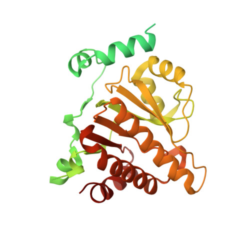
Multiple crystal forms of human MacroD2.

Wazir, S.Maksimainen, M.M.Lehtio, L.

(2020) Acta Crystallogr F Struct Biol Commun 76: 477-482

  • DOI: https://doi.org/10.1107/S2053230X20011309
  • Primary Citation of Related Structures:  
    6Y4Y, 6Y4Z, 6Y73

  • PubMed Abstract: 

    MacroD2 is one of the three human macrodomain proteins characterized by their protein-linked mono-ADP-ribosyl-hydrolyzing activity. MacroD2 is a single-domain protein that contains a deep ADP-ribose-binding groove. In this study, new crystallization conditions for MacroD2 were found and three crystal structures of human MacroD2 in the apo state were solved in space groups P4 1 2 1 2, P4 3 2 1 2 and P4 3 , and refined at 1.75, 1.90 and 1.70 Å resolution, respectively. Structural comparison of the apo crystal structures with the previously reported crystal structure of MacroD2 in complex with ADP-ribose revealed conformational changes in the side chains of Val101, Ile189 and Phe224 induced by the binding of ADP-ribose in the active site. These conformational variations may potentially facilitate design efforts of a MacroD2 inhibitor.


  • Organizational Affiliation

    Faculty of Biochemistry and Molecular Medicine, University of Oulu, PO Box 5400, Oulu 90014, Finland.


Macromolecules
Find similar proteins by:  (by identity cutoff)  |  3D Structure
Entity ID: 1
MoleculeChains Sequence LengthOrganismDetailsImage
ADP-ribose glycohydrolase MACROD2
A, B, C, D, E
A, B, C, D, E, F, G, H
366Homo sapiensMutation(s): 0 
Gene Names: MACROD2C20orf133
EC: 3.5.1 (PDB Primary Data), 3.2.2 (PDB Primary Data)
UniProt & NIH Common Fund Data Resources
Find proteins for A1Z1Q3 (Homo sapiens)
Explore A1Z1Q3 
Go to UniProtKB:  A1Z1Q3
PHAROS:  A1Z1Q3
GTEx:  ENSG00000172264 
Entity Groups  
Sequence Clusters30% Identity50% Identity70% Identity90% Identity95% Identity100% Identity
UniProt GroupA1Z1Q3
Sequence Annotations
Expand
  • Reference Sequence
Small Molecules
Ligands 3 Unique
IDChains Name / Formula / InChI Key2D Diagram3D Interactions
TLA
Query on TLA

Download Ideal Coordinates CCD File 
K [auth B],
L [auth C],
Q [auth F],
S [auth G],
T [auth H]
L(+)-TARTARIC ACID
C4 H6 O6
FEWJPZIEWOKRBE-JCYAYHJZSA-N
GOL
Query on GOL

Download Ideal Coordinates CCD File 
I [auth A],
J [auth B],
M [auth C],
O [auth C],
P [auth D]
GLYCEROL
C3 H8 O3
PEDCQBHIVMGVHV-UHFFFAOYSA-N
DMS
Query on DMS

Download Ideal Coordinates CCD File 
N [auth C],
R [auth F]
DIMETHYL SULFOXIDE
C2 H6 O S
IAZDPXIOMUYVGZ-UHFFFAOYSA-N
Experimental Data & Validation

Experimental Data

  • Method: X-RAY DIFFRACTION
  • Resolution: 1.70 Å
  • R-Value Free: 0.203 
  • R-Value Work: 0.182 
  • R-Value Observed: 0.183 
  • Space Group: P 43
Unit Cell:
Length ( Å )Angle ( ˚ )
a = 96.78α = 90
b = 96.78β = 90
c = 261.09γ = 90
Software Package:
Software NamePurpose
XDSdata reduction
XSCALEdata scaling
REFMACrefinement
PDB_EXTRACTdata extraction
MOLREPphasing

Structure Validation

View Full Validation Report


Entry History & Funding Information

Deposition Data


Funding OrganizationLocationGrant Number
Academy of FinlandFinland287063
Academy of FinlandFinland294085

Revision History  (Full details and data files)

  • Version 1.0: 2020-09-30
    Type: Initial release
  • Version 1.1: 2020-10-14
    Changes: Database references
  • Version 1.2: 2024-01-24
    Changes: Data collection, Database references, Refinement description