6VHQ

Crystal structure of Bacillus subtilis levansucrase (D86A/E342A) in complex with oligosaccharides


Experimental Data Snapshot

  • Method: X-RAY DIFFRACTION
  • Resolution: 2.05 Å
  • R-Value Free: 0.235 
  • R-Value Work: 0.167 
  • R-Value Observed: 0.170 

wwPDB Validation   3D Report Full Report

Currently 6VHQ does not have a validation slider image.


This is version 1.3 of the entry. See complete history


Literature

The molecular basis of the nonprocessive elongation mechanism in levansucrases.

Raga-Carbajal, E.Diaz-Vilchis, A.Rojas-Trejo, S.P.Rudino-Pinera, E.Olvera, C.

(2020) J Biol Chem 296: 100178-100178

  • DOI: https://doi.org/10.1074/jbc.RA120.015853
  • Primary Citation of Related Structures:  
    6VHQ

  • PubMed Abstract: 

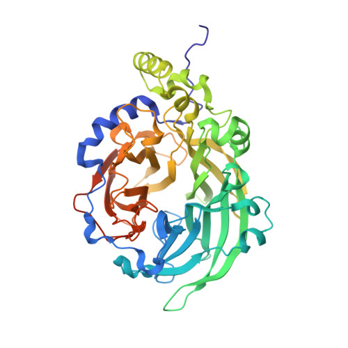
    Levansucrases (LSs) synthesize levan, a β2-6-linked fructose polymer, by successively transferring the fructosyl moiety from sucrose to a growing acceptor molecule. Elucidation of the levan polymerization mechanism is important for using LSs in the production of size-defined products for application in the food and pharmaceutical industries. For a deeper understanding of the levan synthesis reaction, we determined the crystallographic structure of Bacillus subtilis LS (SacB) in complex with a levan-type fructooligosaccharide and utilized site-directed mutagenesis to identify residues involved in substrate binding. The presence of a levanhexaose molecule in the central catalytic cavity allowed us to identify five substrate-binding subsites (-1, +1, +2, +3, and +4). Mutants affecting residues belonging to the identified acceptor subsites showed similar substrate affinity (Km) values to the wildtype (WT) Km value but had a lower turnover number and transfructosylation/hydrolysis ratio. Of importance, compared with the WT, the variants progressively yielded smaller-sized low-molecular-weight levans, as the affected subsites that were closer to the catalytic site, but without affecting their ability to synthesized high-molecular-weight levans. Furthermore, an additional oligosaccharide-binding site 20 Å away from the catalytic pocket was identified, and its potential participation in the elongation mechanism is discussed. Our results clarify, for the first time, the interaction of the enzyme with an acceptor/product oligosaccharide and elucidate the molecular basis of the nonprocessive levan elongation mechanism of LSs.


  • Organizational Affiliation

    Departamento de Ingeniería Celular y Biocatálisis, Instituto de Biotecnología, Universidad Nacional Autónoma de México, Cuernavaca, Morelos, México.


Macromolecules
Find similar proteins by:  (by identity cutoff)  |  3D Structure
Entity ID: 1
MoleculeChains Sequence LengthOrganismDetailsImage
Glycoside hydrolase family 68 protein
A, B
466Bacillus subtilisMutation(s): 2 
Gene Names: sacBB4417_3269ETA10_18085ETL41_09350FVD40_16900
UniProt
Find proteins for P05655 (Bacillus subtilis (strain 168))
Explore P05655 
Go to UniProtKB:  P05655
Entity Groups  
Sequence Clusters30% Identity50% Identity70% Identity90% Identity95% Identity100% Identity
UniProt GroupP05655
Sequence Annotations
Expand
  • Reference Sequence
Oligosaccharides

Help

Entity ID: 2
MoleculeChains Length2D Diagram Glycosylation3D Interactions
beta-D-fructofuranose-(2-6)-beta-D-fructofuranose-(2-6)-beta-D-fructofuranose-(2-6)-beta-D-fructofuranose-(2-6)-beta-D-fructofuranose-(2-6)-beta-D-fructofuranose
C, D, E
6N/A
Glycosylation Resources
GlyTouCan:  G00825VL
GlyCosmos:  G00825VL
Entity ID: 3
MoleculeChains Length2D Diagram Glycosylation3D Interactions
beta-D-fructofuranose-(2-6)-beta-D-fructofuranose
F
2N/A
Glycosylation Resources
GlyTouCan:  G51829YA
GlyCosmos:  G51829YA
Small Molecules
Ligands 2 Unique
IDChains Name / Formula / InChI Key2D Diagram3D Interactions
BR (Subject of Investigation/LOI)
Query on BR

Download Ideal Coordinates CCD File 
H [auth A]
I [auth A]
J [auth A]
K [auth A]
L [auth A]
H [auth A],
I [auth A],
J [auth A],
K [auth A],
L [auth A],
M [auth A],
O [auth B],
P [auth B],
Q [auth B]
BROMIDE ION
Br
CPELXLSAUQHCOX-UHFFFAOYSA-M
CA (Subject of Investigation/LOI)
Query on CA

Download Ideal Coordinates CCD File 
G [auth A],
N [auth B]
CALCIUM ION
Ca
BHPQYMZQTOCNFJ-UHFFFAOYSA-N
Biologically Interesting Molecules (External Reference) 1 Unique
Experimental Data & Validation

Experimental Data

  • Method: X-RAY DIFFRACTION
  • Resolution: 2.05 Å
  • R-Value Free: 0.235 
  • R-Value Work: 0.167 
  • R-Value Observed: 0.170 
  • Space Group: P 1 21 1
Unit Cell:
Length ( Å )Angle ( ˚ )
a = 69.313α = 90
b = 78.665β = 93.94
c = 78.732γ = 90
Software Package:
Software NamePurpose
PHENIXrefinement
PDB_EXTRACTdata extraction
HKL-3000data reduction
HKL-3000data scaling
PHASERphasing

Structure Validation

View Full Validation Report

Currently 6VHQ does not have a validation slider image.


Entry History & Funding Information

Deposition Data


Funding OrganizationLocationGrant Number
Other governmentMexicoConsejo Nacional de Ciencia y Tecnologia-CB-2013-219728
Other governmentMexicoConsejo Nacional de Ciencia y Tecnologia-BMBF-2015-267620
Other governmentMexicoUniversidad Nacional Autonoma de Mexico-PAPIIT IN213616

Revision History  (Full details and data files)

  • Version 1.0: 2021-01-13
    Type: Initial release
  • Version 1.1: 2021-07-14
    Changes: Database references
  • Version 1.2: 2021-07-21
    Changes: Database references
  • Version 1.3: 2023-10-11
    Changes: Data collection, Database references, Refinement description