6TV0

Serratia spp. cyanase hydratase


Experimental Data Snapshot

  • Method: X-RAY DIFFRACTION
  • Resolution: 1.96 Å
  • R-Value Free: 0.218 
  • R-Value Work: 0.176 
  • R-Value Observed: 0.178 

wwPDB Validation   3D Report Full Report


Ligand Structure Quality Assessment 


This is version 1.2 of the entry. See complete history


Literature

Detecting the nature and solving the crystal structure of a contaminant protein from an opportunistic pathogen.

Pederzoli, R.Tarantino, D.Gourlay, L.J.Chaves-Sanjuan, A.Bolognesi, M.

(2020) Acta Crystallogr F Struct Biol Commun 76: 392-397

  • DOI: https://doi.org/10.1107/S2053230X20010626
  • Primary Citation of Related Structures:  
    6TV0

  • PubMed Abstract: 

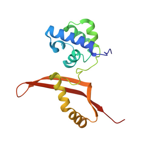
    The unintentional crystallization of contaminant proteins in the place of target recombinant proteins is sporadically reported, despite the availability of stringent expression/purification protocols and of software for the detection of contaminants. Typically, the contaminant protein originates from the expression organism (for example Escherichia coli), but in rare circumstances contaminants from different sources have been reported. Here, a case of contamination from a Serratia bacterial strain that occurred while attempting to crystallize an unrelated protein from Burkholderia pseudomallei (overexpressed in E. coli) is presented. The contamination led to the unintended crystallization and structure analysis of a cyanase hydratase from a bacterial strain of the Serratia genus, an opportunistic enterobacterium that grows under conditions similar to those of E. coli and that is found in a variety of habitats, including the laboratory environment. In this context, the procedures that were adopted to identify the contaminant based on crystallographic data only are presented and the crystal structure of Serrata spp. cyanase hydratase is briefly discussed.


  • Organizational Affiliation

    Hamburg Unit c/o DESY, European Molecular Biology Laboratory, Notkestrasse 85, 22603 Hamburg, Germany.


Macromolecules
Find similar proteins by:  (by identity cutoff)  |  3D Structure
Entity ID: 1
MoleculeChains Sequence LengthOrganismDetailsImage
Cyanate hydratase
A, B, C, D, E
A, B, C, D, E, F, G, H, I, J
155Serratia sp. (in: enterobacteria)Mutation(s): 0 
EC: 4.2.1.104
Entity Groups  
Sequence Clusters30% Identity50% Identity70% Identity90% Identity95% Identity100% Identity
Sequence Annotations
Expand
  • Reference Sequence
Small Molecules
Ligands 2 Unique
IDChains Name / Formula / InChI Key2D Diagram3D Interactions
GOL (Subject of Investigation/LOI)
Query on GOL

Download Ideal Coordinates CCD File 
AA [auth C]
AB [auth H]
BA [auth C]
BB [auth H]
CA [auth C]
AA [auth C],
AB [auth H],
BA [auth C],
BB [auth H],
CA [auth C],
CB [auth H],
DA [auth C],
DB [auth H],
EA [auth D],
EB [auth H],
FA [auth D],
FB [auth H],
GA [auth D],
GB [auth I],
HA [auth D],
HB [auth I],
IA [auth D],
IB [auth I],
JA [auth D],
JB [auth J],
KA [auth E],
KB [auth J],
L [auth A],
LA [auth E],
LB [auth J],
M [auth A],
MA [auth E],
MB [auth J],
N [auth A],
NA [auth E],
O [auth A],
OA [auth E],
P [auth A],
PA [auth E],
RA [auth F],
S [auth B],
SA [auth F],
T [auth B],
TA [auth F],
U [auth B],
UA [auth G],
V [auth B],
VA [auth G],
W [auth B],
WA [auth G],
X [auth B],
XA [auth G],
YA [auth G],
Z [auth C],
ZA [auth G]
GLYCEROL
C3 H8 O3
PEDCQBHIVMGVHV-UHFFFAOYSA-N
OXD (Subject of Investigation/LOI)
Query on OXD

Download Ideal Coordinates CCD File 
K [auth A],
Q [auth A],
QA [auth F],
R [auth B],
Y [auth C]
OXALIC ACID
C2 H2 O4
MUBZPKHOEPUJKR-UHFFFAOYSA-N
Experimental Data & Validation

Experimental Data

  • Method: X-RAY DIFFRACTION
  • Resolution: 1.96 Å
  • R-Value Free: 0.218 
  • R-Value Work: 0.176 
  • R-Value Observed: 0.178 
  • Space Group: P 1 21 1
Unit Cell:
Length ( Å )Angle ( ˚ )
a = 86.793α = 90
b = 93.14β = 95.743
c = 111.193γ = 90
Software Package:
Software NamePurpose
REFMACrefinement
PHENIXrefinement
XDSdata reduction
Aimlessdata scaling
MOLREPphasing
PHASERphasing

Structure Validation

View Full Validation Report


Ligand Structure Quality Assessment 


Entry History 

Deposition Data

Revision History  (Full details and data files)

  • Version 1.0: 2021-01-27
    Type: Initial release
  • Version 1.1: 2022-02-09
    Changes: Database references
  • Version 1.2: 2024-01-24
    Changes: Data collection, Refinement description