6TT4

Crystal structure of 'Res_S2 mutant human Angiotensin-1 converting enzyme N-domain in complex with omapatrilat.


Experimental Data Snapshot

  • Method: X-RAY DIFFRACTION
  • Resolution: 1.80 Å
  • R-Value Free: 0.222 
  • R-Value Work: 0.182 
  • R-Value Observed: 0.182 

wwPDB Validation   3D Report Full Report


Ligand Structure Quality Assessment 


This is version 2.1 of the entry. See complete history


Literature

ACE-domain selectivity extends beyond direct interacting residues at the active site.

Cozier, G.E.Lubbe, L.Sturrock, E.D.Acharya, K.R.

(2020) Biochem J 477: 1241-1259

  • DOI: https://doi.org/10.1042/BCJ20200060
  • Primary Citation of Related Structures:  
    6TT1, 6TT3, 6TT4

  • PubMed Abstract: 

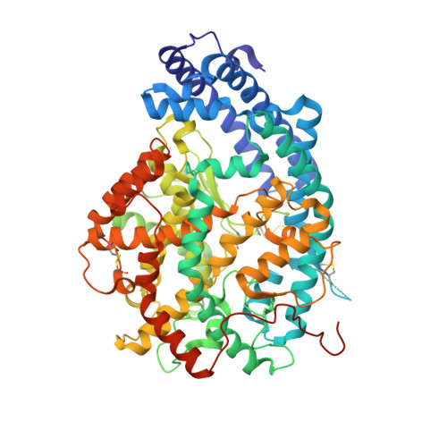
    Angiotensin-converting enzyme (ACE) is best known for its formation of the vasopressor angiotensin II that controls blood pressure but is also involved in other physiological functions through the hydrolysis of a variety of peptide substrates. The enzyme contains two catalytic domains (nACE and cACE) that have different affinities for ACE substrates and inhibitors. We investigated whether nACE inhibitor backbones contain a unique property which allows them to take advantage of the hinging of nACE. Kinetic analysis showed that mutation of unique nACE residues, in both the S2 pocket and around the prime subsites (S') to their C-domain counterparts, each resulted in a decrease in the affinity of nACE specific inhibitors (SG6, 33RE and ketoACE-13) but it required the combined S2_S' mutant to abrogate nACE-selectivity. However, this was not observed with the non-domain-selective inhibitors enalaprilat and omapatrilat. High-resolution structures were determined for the minimally glycosylated nACE with the combined S2_S' mutations in complex with the ACE inhibitors 33RE (1.8 Å), omapatrilat (1.8 Å) and SG6 (1.7 Å). These confirmed that the affinities of the nACE-selective SG6, 33RE and ketoACE-13 are not only affected by direct interactions with the immediate environment of the binding site, but also by more distal residues. This study provides evidence for a more general mechanism of ACE inhibition involving synergistic effects of not only the S2, S1' and S2' subsites, but also residues involved in the sub-domain interface that effect the unique ways in which the two domains stabilize active site loops to favour inhibitor binding.


  • Organizational Affiliation

    Department of Biology and Biochemistry, University of Bath, Claverton Down, Bath BA2 7AY, U.K.


Macromolecules
Find similar proteins by:  (by identity cutoff)  |  3D Structure
Entity ID: 1
MoleculeChains Sequence LengthOrganismDetailsImage
Angiotensin-converting enzyme
A, B
629Homo sapiensMutation(s): 0 
Gene Names: ACEDCPDCP1
EC: 3.2.1 (PDB Primary Data), 3.4.15.1 (PDB Primary Data)
UniProt & NIH Common Fund Data Resources
Find proteins for P12821 (Homo sapiens)
Explore P12821 
Go to UniProtKB:  P12821
PHAROS:  P12821
GTEx:  ENSG00000159640 
Entity Groups  
Sequence Clusters30% Identity50% Identity70% Identity90% Identity95% Identity100% Identity
UniProt GroupP12821
Sequence Annotations
Expand
  • Reference Sequence
Oligosaccharides

Help

Entity ID: 2
MoleculeChains Length2D Diagram Glycosylation3D Interactions
beta-D-mannopyranose-(1-4)-2-acetamido-2-deoxy-beta-D-glucopyranose-(1-4)-2-acetamido-2-deoxy-beta-D-glucopyranose
C
3N-Glycosylation
Glycosylation Resources
GlyTouCan:  G15407YE
GlyCosmos:  G15407YE
GlyGen:  G15407YE
Entity ID: 3
MoleculeChains Length2D Diagram Glycosylation3D Interactions
2-acetamido-2-deoxy-beta-D-glucopyranose-(1-4)-[alpha-L-fucopyranose-(1-6)]2-acetamido-2-deoxy-beta-D-glucopyranose
D
3N-Glycosylation
Glycosylation Resources
GlyTouCan:  G21290RB
GlyCosmos:  G21290RB
GlyGen:  G21290RB
Entity ID: 4
MoleculeChains Length2D Diagram Glycosylation3D Interactions
2-acetamido-2-deoxy-beta-D-glucopyranose-(1-4)-2-acetamido-2-deoxy-beta-D-glucopyranose
E
2N-Glycosylation
Glycosylation Resources
GlyTouCan:  G42666HT
GlyCosmos:  G42666HT
GlyGen:  G42666HT
Entity ID: 5
MoleculeChains Length2D Diagram Glycosylation3D Interactions
alpha-D-mannopyranose-(1-3)-beta-D-mannopyranose-(1-4)-2-acetamido-2-deoxy-beta-D-glucopyranose-(1-4)-[alpha-L-fucopyranose-(1-6)]2-acetamido-2-deoxy-beta-D-glucopyranose
F
5N-Glycosylation
Glycosylation Resources
GlyTouCan:  G42466VF
GlyCosmos:  G42466VF
GlyGen:  G42466VF
Entity ID: 6
MoleculeChains Length2D Diagram Glycosylation3D Interactions
alpha-L-fucopyranose-(1-6)-2-acetamido-2-deoxy-beta-D-glucopyranose
G
2N-Glycosylation
Glycosylation Resources
GlyTouCan:  G86851RC
GlyCosmos:  G86851RC
GlyGen:  G86851RC
Small Molecules
Ligands 10 Unique
IDChains Name / Formula / InChI Key2D Diagram3D Interactions
XPE
Query on XPE

Download Ideal Coordinates CCD File 
L [auth A]3,6,9,12,15,18,21,24,27-NONAOXANONACOSANE-1,29-DIOL
C20 H42 O11
DTPCFIHYWYONMD-UHFFFAOYSA-N
FT8 (Subject of Investigation/LOI)
Query on FT8

Download Ideal Coordinates CCD File 
J [auth A],
U [auth B]
Omapatrilat
C19 H24 N2 O4 S2
LVRLSYPNFFBYCZ-VGWMRTNUSA-N
NAG
Query on NAG

Download Ideal Coordinates CCD File 
H [auth A]2-acetamido-2-deoxy-beta-D-glucopyranose
C8 H15 N O6
OVRNDRQMDRJTHS-FMDGEEDCSA-N
BCN
Query on BCN

Download Ideal Coordinates CCD File 
BA [auth B],
Q [auth A]
BICINE
C6 H13 N O4
FSVCELGFZIQNCK-UHFFFAOYSA-N
PGE
Query on PGE

Download Ideal Coordinates CCD File 
M [auth A],
W [auth B]
TRIETHYLENE GLYCOL
C6 H14 O4
ZIBGPFATKBEMQZ-UHFFFAOYSA-N
PEG
Query on PEG

Download Ideal Coordinates CCD File 
P [auth A]DI(HYDROXYETHYL)ETHER
C4 H10 O3
MTHSVFCYNBDYFN-UHFFFAOYSA-N
ZN
Query on ZN

Download Ideal Coordinates CCD File 
I [auth A],
T [auth B]
ZINC ION
Zn
PTFCDOFLOPIGGS-UHFFFAOYSA-N
EDO
Query on EDO

Download Ideal Coordinates CCD File 
AA [auth B]
N [auth A]
O [auth A]
X [auth B]
Y [auth B]
AA [auth B],
N [auth A],
O [auth A],
X [auth B],
Y [auth B],
Z [auth B]
1,2-ETHANEDIOL
C2 H6 O2
LYCAIKOWRPUZTN-UHFFFAOYSA-N
BO3
Query on BO3

Download Ideal Coordinates CCD File 
CA [auth B],
DA [auth B],
R [auth A],
S [auth A]
BORIC ACID
B H3 O3
KGBXLFKZBHKPEV-UHFFFAOYSA-N
CL
Query on CL

Download Ideal Coordinates CCD File 
K [auth A],
V [auth B]
CHLORIDE ION
Cl
VEXZGXHMUGYJMC-UHFFFAOYSA-M
Experimental Data & Validation

Experimental Data

  • Method: X-RAY DIFFRACTION
  • Resolution: 1.80 Å
  • R-Value Free: 0.222 
  • R-Value Work: 0.182 
  • R-Value Observed: 0.182 
  • Space Group: P 1
Unit Cell:
Length ( Å )Angle ( ˚ )
a = 74.371α = 92.433
b = 78.825β = 105.966
c = 89.686γ = 114.603
Software Package:
Software NamePurpose
PHENIXrefinement
PHENIXrefinement
DIALSdata reduction
Aimlessdata scaling
PHASERphasing

Structure Validation

View Full Validation Report


Ligand Structure Quality Assessment 


Entry History & Funding Information

Deposition Data


Funding OrganizationLocationGrant Number
Medical Research Council (MRC, United Kingdom)United KingdomMR/M026647/1

Revision History  (Full details and data files)

  • Version 1.0: 2020-04-01
    Type: Initial release
  • Version 1.1: 2020-04-22
    Changes: Database references
  • Version 2.0: 2020-07-29
    Type: Remediation
    Reason: Carbohydrate remediation
    Changes: Atomic model, Data collection, Derived calculations, Structure summary
  • Version 2.1: 2024-01-24
    Changes: Data collection, Database references, Derived calculations, Refinement description, Structure summary