6TD2

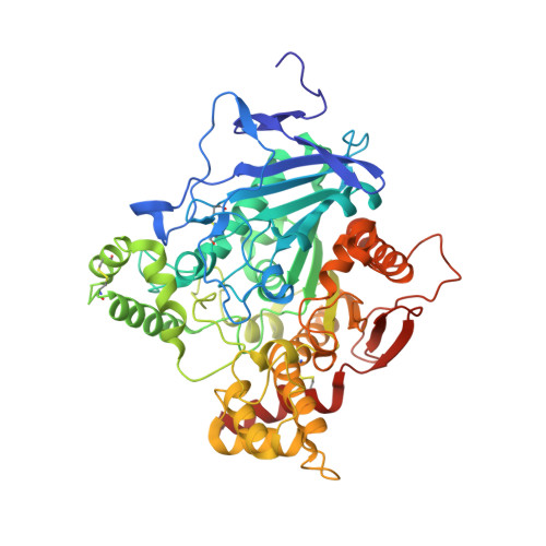
Mus musculus Acetylcholinesterase in complex with N-(2-(diethylamino)ethyl)-1-(4-(trifluoromethyl)phenyl)methanesulfonamide


Experimental Data Snapshot

  • Method: X-RAY DIFFRACTION
  • Resolution: 2.80 Å
  • R-Value Free: 0.215 
  • R-Value Work: 0.175 
  • R-Value Observed: 0.175 

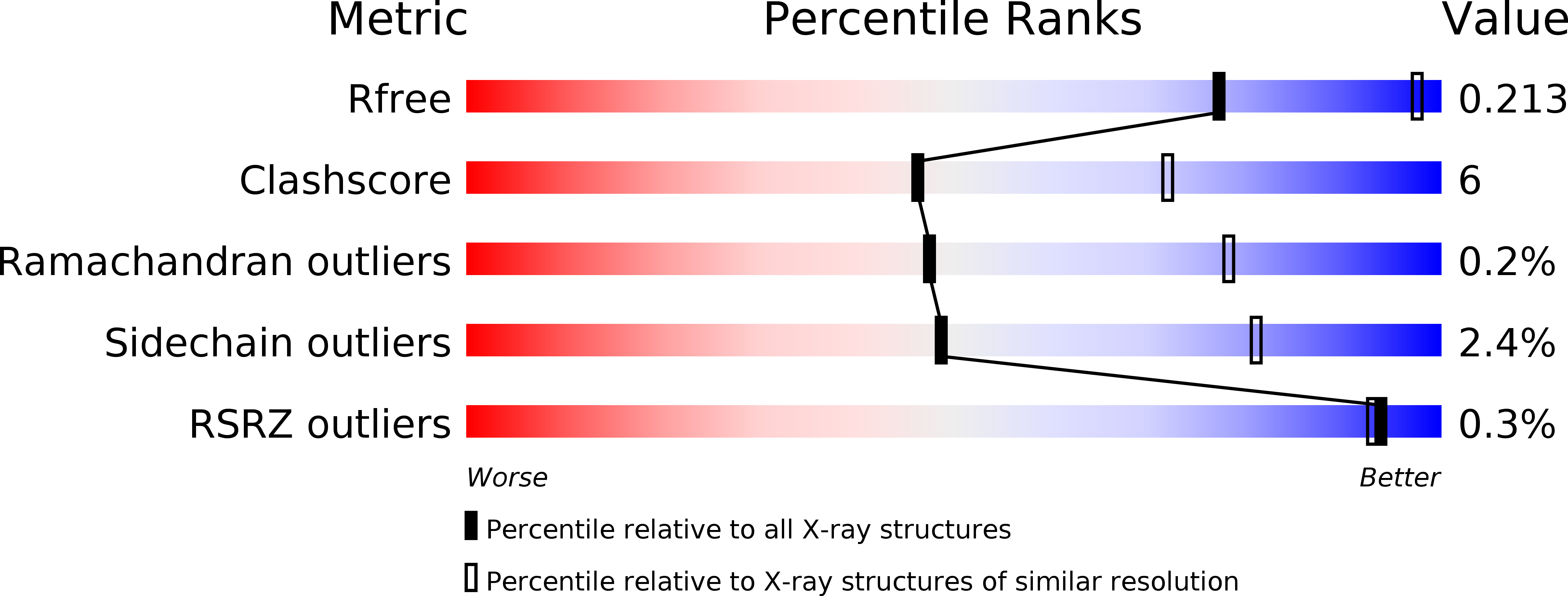
wwPDB Validation   3D Report Full Report


Ligand Structure Quality Assessment 


This is version 1.2 of the entry. See complete history


Literature

Physical Mechanisms Governing Substituent Effects on Arene-Arene Interactions in a Protein Milieu.

Andersson, C.D.Mishra, B.K.Forsgren, N.Ekstrom, F.Linusson, A.

(2020) J Phys Chem B 124: 6529-6539

  • DOI: https://doi.org/10.1021/acs.jpcb.0c03778
  • Primary Citation of Related Structures:  
    6TD2

  • PubMed Abstract: 

    Arene-arene interactions play important roles in protein-ligand complex formation. Here, we investigate the characteristics of arene-arene interactions between small organic molecules and aromatic amino acids in protein interiors. The study is based on X-ray crystallographic data and quantum mechanical calculations using the enzyme acetylcholinesterase and selected inhibitory ligands as a model system. It is shown that the arene substituents of the inhibitors dictate the strength of the interaction and the geometry of the resulting complexes. Importantly, the calculated interaction energies correlate well with the measured inhibitor potency. Non-hydrogen substituents strengthened all interaction types in the protein milieu, in keeping with results for benzene dimer model systems. The interaction energies were dispersion-dominated, but substituents that induced local dipole moments increased the electrostatic contribution and thus yielded more strongly bound complexes. These findings provide fundamental insights into the physical mechanisms governing arene-arene interactions in the protein milieu and thus into molecular recognition between proteins and small molecules.


  • Organizational Affiliation

    Department of Chemistry, Umeå University, SE-90187 Umeå, Sweden.


Macromolecules
Find similar proteins by:  (by identity cutoff)  |  3D Structure
Entity ID: 1
MoleculeChains Sequence LengthOrganismDetailsImage
Acetylcholinesterase
A, B
543Mus musculusMutation(s): 0 
Gene Names: Ache
EC: 3.1.1.7
UniProt & NIH Common Fund Data Resources
Find proteins for P21836 (Mus musculus)
Explore P21836 
Go to UniProtKB:  P21836
IMPC:  MGI:87876
Entity Groups  
Sequence Clusters30% Identity50% Identity70% Identity90% Identity95% Identity100% Identity
UniProt GroupP21836
Sequence Annotations
Expand
  • Reference Sequence
Small Molecules
Ligands 6 Unique
IDChains Name / Formula / InChI Key2D Diagram3D Interactions
PE4
Query on PE4

Download Ideal Coordinates CCD File 
L [auth A]2-{2-[2-(2-{2-[2-(2-ETHOXY-ETHOXY)-ETHOXY]-ETHOXY}-ETHOXY)-ETHOXY]-ETHOXY}-ETHANOL
C16 H34 O8
PJWQOENWHPEPKI-UHFFFAOYSA-N
N2K (Subject of Investigation/LOI)
Query on N2K

Download Ideal Coordinates CCD File 
E [auth A],
M [auth B]
~{N}-[2-(diethylamino)ethyl]-1-[4-(trifluoromethyl)phenyl]methanesulfonamide
C14 H21 F3 N2 O2 S
UJMFKSSVUDLNIV-UHFFFAOYSA-N
NAG
Query on NAG

Download Ideal Coordinates CCD File 
C [auth A],
D [auth A]
2-acetamido-2-deoxy-beta-D-glucopyranose
C8 H15 N O6
OVRNDRQMDRJTHS-FMDGEEDCSA-N
ETE
Query on ETE

Download Ideal Coordinates CCD File 
S [auth B]2-{2-[2-2-(METHOXY-ETHOXY)-ETHOXY]-ETHOXY}-ETHANOL
C9 H20 O5
ZNYRFEPBTVGZDN-UHFFFAOYSA-N
PG0
Query on PG0

Download Ideal Coordinates CCD File 
I [auth A],
J [auth A],
K [auth A],
Q [auth B],
R [auth B]
2-(2-METHOXYETHOXY)ETHANOL
C5 H12 O3
SBASXUCJHJRPEV-UHFFFAOYSA-N
EDO
Query on EDO

Download Ideal Coordinates CCD File 
F [auth A]
G [auth A]
H [auth A]
N [auth B]
O [auth B]
F [auth A],
G [auth A],
H [auth A],
N [auth B],
O [auth B],
P [auth B]
1,2-ETHANEDIOL
C2 H6 O2
LYCAIKOWRPUZTN-UHFFFAOYSA-N
Experimental Data & Validation

Experimental Data

  • Method: X-RAY DIFFRACTION
  • Resolution: 2.80 Å
  • R-Value Free: 0.215 
  • R-Value Work: 0.175 
  • R-Value Observed: 0.175 
  • Space Group: P 21 21 21
Unit Cell:
Length ( Å )Angle ( ˚ )
a = 78.957α = 90
b = 111.957β = 90
c = 227.429γ = 90
Software Package:
Software NamePurpose
PHENIXrefinement
XDSdata reduction
SCALAdata scaling
REFMACphasing

Structure Validation

View Full Validation Report


Ligand Structure Quality Assessment 


Entry History & Funding Information

Deposition Data


Funding OrganizationLocationGrant Number
Other government--
Swedish Research Council--

Revision History  (Full details and data files)

  • Version 1.0: 2020-10-14
    Type: Initial release
  • Version 1.1: 2020-10-21
    Changes: Derived calculations
  • Version 1.2: 2024-01-24
    Changes: Data collection, Database references, Refinement description