6RYO

Bacterial membrane enzyme structure by the in meso method at 1.9 A resolution


Experimental Data Snapshot

  • Method: X-RAY DIFFRACTION
  • Resolution: 1.92 Å
  • R-Value Free: 0.276 
  • R-Value Work: 0.250 
  • R-Value Observed: 0.252 

wwPDB Validation   3D Report Full Report


Ligand Structure Quality Assessment 


This is version 2.1 of the entry. See complete history


Literature

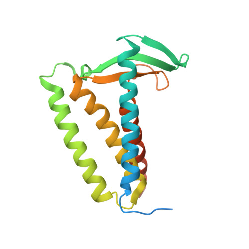
Structures of lipoprotein signal peptidase II from Staphylococcus aureus complexed with antibiotics globomycin and myxovirescin.

Olatunji, S.Yu, X.Bailey, J.Huang, C.Y.Zapotoczna, M.Bowen, K.Remskar, M.Muller, R.Scanlan, E.M.Geoghegan, J.A.Olieric, V.Caffrey, M.

(2020) Nat Commun 11: 140-140

  • DOI: https://doi.org/10.1038/s41467-019-13724-y
  • Primary Citation of Related Structures:  
    6RYO, 6RYP

  • PubMed Abstract: 

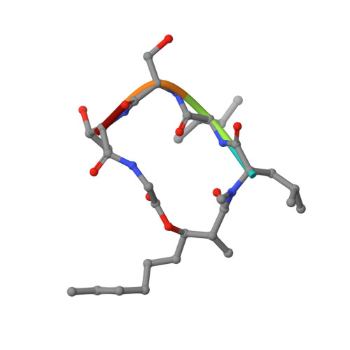
    Antimicrobial resistance is a major global threat that calls for new antibiotics. Globomycin and myxovirescin are two natural antibiotics that target the lipoprotein-processing enzyme, LspA, thereby compromising the integrity of the bacterial cell envelope. As part of a project aimed at understanding their mechanism of action and for drug development, we provide high-resolution crystal structures of the enzyme from the human pathogen methicillin-resistant Staphylococcus aureus (MRSA) complexed with globomycin and with myxovirescin. Our results reveal an instance of convergent evolution. The two antibiotics possess different molecular structures. Yet, they appear to inhibit identically as non-cleavable tetrahedral intermediate analogs. Remarkably, the two antibiotics superpose along nineteen contiguous atoms that interact similarly with LspA. This 19-atom motif recapitulates a part of the substrate lipoprotein in its proposed binding mode. Incorporating this motif into a scaffold with suitable pharmacokinetic properties should enable the development of effective antibiotics with built-in resistance hardiness.


  • Organizational Affiliation

    Membrane Structural and Functional Biology Group, School of Medicine and School of Biochemistry and Immunology, Trinity College Dublin, Dublin, D02 R590, Ireland.


Macromolecules
Find similar proteins by:  (by identity cutoff)  |  3D Structure
Entity ID: 1
MoleculeChains Sequence LengthOrganismDetailsImage
Lipoprotein signal peptidase187Staphylococcus aureusMutation(s): 0 
Gene Names: lspAlspSAR1172
EC: 3.4.23.36
Membrane Entity: Yes 
UniProt
Find proteins for Q6GHN9 (Staphylococcus aureus (strain MRSA252))
Explore Q6GHN9 
Go to UniProtKB:  Q6GHN9
Entity Groups  
Sequence Clusters30% Identity50% Identity70% Identity90% Identity95% Identity100% Identity
UniProt GroupQ6GHN9
Sequence Annotations
Expand
  • Reference Sequence

Find similar proteins by:  Sequence   |   3D Structure  

Entity ID: 2
MoleculeChains Sequence LengthOrganismDetailsImage
Globomycin5synthetic constructMutation(s): 0 
Sequence Annotations
Expand
  • Reference Sequence
Small Molecules
Ligands 4 Unique
IDChains Name / Formula / InChI Key2D Diagram3D Interactions
OLC
Query on OLC

Download Ideal Coordinates CCD File 
C [auth A]
D [auth A]
E [auth A]
F [auth A]
G [auth A]
C [auth A],
D [auth A],
E [auth A],
F [auth A],
G [auth A],
W [auth B]
(2R)-2,3-dihydroxypropyl (9Z)-octadec-9-enoate
C21 H40 O4
RZRNAYUHWVFMIP-GDCKJWNLSA-N
7PE
Query on 7PE

Download Ideal Coordinates CCD File 
K [auth A]
L [auth A]
M [auth A]
N [auth A]
O [auth A]
K [auth A],
L [auth A],
M [auth A],
N [auth A],
O [auth A],
P [auth A],
Q [auth A],
R [auth A],
T [auth A],
X [auth B]
2-(2-(2-(2-(2-(2-ETHOXYETHOXY)ETHOXY)ETHOXY)ETHOXY)ETHOXY)ETHANOL
C14 H30 O7
UKXKPKBTMYNOFS-UHFFFAOYSA-N
GOL
Query on GOL

Download Ideal Coordinates CCD File 
H [auth A],
I [auth A],
J [auth A],
U [auth A],
V [auth A]
GLYCEROL
C3 H8 O3
PEDCQBHIVMGVHV-UHFFFAOYSA-N
ZN
Query on ZN

Download Ideal Coordinates CCD File 
S [auth A]ZINC ION
Zn
PTFCDOFLOPIGGS-UHFFFAOYSA-N
Modified Residues  3 Unique
IDChains TypeFormula2D DiagramParent
ALO
Query on ALO
B
L-PEPTIDE LINKINGC4 H9 N O3THR
IIL
Query on IIL
B
L-PEPTIDE LINKINGC6 H13 N O2ILE
MLE
Query on MLE
B
L-PEPTIDE LINKINGC7 H15 N O2LEU
Experimental Data & Validation

Experimental Data

  • Method: X-RAY DIFFRACTION
  • Resolution: 1.92 Å
  • R-Value Free: 0.276 
  • R-Value Work: 0.250 
  • R-Value Observed: 0.252 
  • Space Group: P 32 2 1
Unit Cell:
Length ( Å )Angle ( ˚ )
a = 52.258α = 90
b = 52.258β = 90
c = 135.882γ = 120
Software Package:
Software NamePurpose
PHENIXrefinement
autoPROCdata reduction
XSCALEdata scaling
PHASERphasing

Structure Validation

View Full Validation Report


Ligand Structure Quality Assessment 


Entry History & Funding Information

Deposition Data


Funding OrganizationLocationGrant Number
Science Foundation IrelandIreland16/IA/4435
European UnionSwitzerland701647

Revision History  (Full details and data files)

  • Version 1.0: 2020-01-15
    Type: Initial release
  • Version 1.1: 2020-01-22
    Changes: Database references
  • Version 2.0: 2023-11-15
    Changes: Advisory, Atomic model, Data collection, Database references, Derived calculations, Refinement description
  • Version 2.1: 2024-01-24
    Changes: Refinement description