6MRI

E. coli cysteine desulfurase SufS E250A with a cysteine persulfide intermediate


Experimental Data Snapshot

  • Method: X-RAY DIFFRACTION
  • Resolution: 2.62 Å
  • R-Value Free: 0.220 
  • R-Value Work: 0.184 
  • R-Value Observed: 0.187 

wwPDB Validation   3D Report Full Report


Ligand Structure Quality Assessment 


This is version 1.4 of the entry. See complete history


Literature

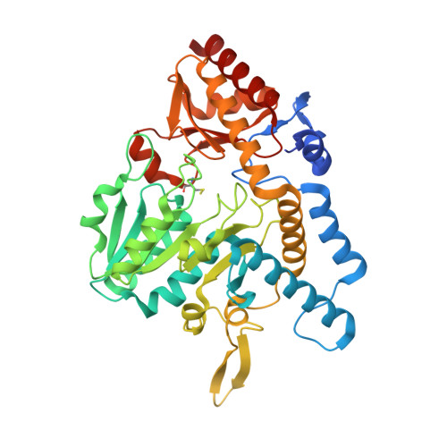
Structural Evidence for Dimer-Interface-Driven Regulation of the Type II Cysteine Desulfurase, SufS.

Dunkle, J.A.Bruno, M.R.Outten, F.W.Frantom, P.A.

(2019) Biochemistry 58: 687-696

  • DOI: https://doi.org/10.1021/acs.biochem.8b01122
  • Primary Citation of Related Structures:  
    6MR2, 6MR6, 6MRE, 6MRH, 6MRI

  • PubMed Abstract: 

    SufS is a type II cysteine desulfurase and acts as the initial step in the Suf Fe-S cluster assembly pathway. In Escherichia coli, this pathway is utilized under conditions of oxidative stress and is resistant to reactive oxygen species. Mechanistically, this means SufS must shift between protecting a covalent persulfide intermediate and making it available for transfer to the next protein partner in the pathway, SufE. Here, we report five X-ray crystal structures of SufS including a new structure of SufS containing an inward-facing persulfide intermediate on C364. Additional structures of SufS variants with substitutions at the dimer interface show changes in dimer geometry and suggest a conserved β-hairpin structure plays a role in mediating interactions with SufE. These new structures, along with previous HDX-MS and biochemical data, identify an interaction network capable of communication between active-sites of the SufS dimer coordinating the shift between desulfurase and transpersulfurase activities.


  • Organizational Affiliation

    Department of Chemistry & Biochemistry , The University of Alabama , Tuscaloosa , Alabama 35487 , United States.


Macromolecules
Find similar proteins by:  (by identity cutoff)  |  3D Structure
Entity ID: 1
MoleculeChains Sequence LengthOrganismDetailsImage
Cysteine desulfurase420Escherichia coli K-12Mutation(s): 1 
Gene Names: sufScsdBynhBb1680JW1670
EC: 2.8.1.7 (PDB Primary Data), 4.4.1.16 (PDB Primary Data)
UniProt
Find proteins for P77444 (Escherichia coli (strain K12))
Explore P77444 
Go to UniProtKB:  P77444
Entity Groups  
Sequence Clusters30% Identity50% Identity70% Identity90% Identity95% Identity100% Identity
UniProt GroupP77444
Sequence Annotations
Expand
  • Reference Sequence
Small Molecules
Ligands 1 Unique
IDChains Name / Formula / InChI Key2D Diagram3D Interactions
PLP
Query on PLP

Download Ideal Coordinates CCD File 
B [auth A]PYRIDOXAL-5'-PHOSPHATE
C8 H10 N O6 P
NGVDGCNFYWLIFO-UHFFFAOYSA-N
Modified Residues  1 Unique
IDChains TypeFormula2D DiagramParent
CSS
Query on CSS
A
L-PEPTIDE LINKINGC3 H7 N O2 S2CYS
Experimental Data & Validation

Experimental Data

  • Method: X-RAY DIFFRACTION
  • Resolution: 2.62 Å
  • R-Value Free: 0.220 
  • R-Value Work: 0.184 
  • R-Value Observed: 0.187 
  • Space Group: P 43 21 2
Unit Cell:
Length ( Å )Angle ( ˚ )
a = 125.73α = 90
b = 125.73β = 90
c = 138.55γ = 90
Software Package:
Software NamePurpose
XSCALEdata scaling
PHASERphasing
PHENIXrefinement
PDB_EXTRACTdata extraction
XDSdata reduction

Structure Validation

View Full Validation Report


Ligand Structure Quality Assessment 


Entry History & Funding Information

Deposition Data


Funding OrganizationLocationGrant Number
National Institutes of Health/National Institute of General Medical Sciences (NIH/NIGMS)United StatesGM112919

Revision History  (Full details and data files)

  • Version 1.0: 2019-01-09
    Type: Initial release
  • Version 1.1: 2019-02-27
    Changes: Data collection, Database references
  • Version 1.2: 2020-01-01
    Changes: Author supporting evidence
  • Version 1.3: 2023-10-11
    Changes: Data collection, Database references, Derived calculations, Refinement description
  • Version 1.4: 2023-11-15
    Changes: Data collection