


Crystal Structure of dUTP pyrophosphatase protein, from Naegleria fowleri in complex with deoxyuridine
Delker, S.L., Abendroth, J., Lorimer, D., Edwards, T.E.To be published.
Experimental Data Snapshot
wwPDB Validation 3D Report Full Report
Currently 6MJK does not have a validation slider image.
Entity ID: 1 | |||||
|---|---|---|---|---|---|
| Molecule | Chains | Sequence Length | Organism | Details | Image |
| dUTP pyrophosphatase | 155 | Naegleria fowleri | Mutation(s): 0 | 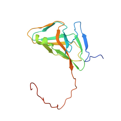 | |
UniProt | |||||
Find proteins for A0A1Z0YU86 (Naegleria fowleri) Explore A0A1Z0YU86 Go to UniProtKB: A0A1Z0YU86 | |||||
Entity Groups | |||||
| Sequence Clusters | 30% Identity50% Identity70% Identity90% Identity95% Identity100% Identity | ||||
| UniProt Group | A0A1Z0YU86 | ||||
Sequence AnnotationsExpand | |||||
| |||||
| Ligands 3 Unique | |||||
|---|---|---|---|---|---|
| ID | Chains | Name / Formula / InChI Key | 2D Diagram | 3D Interactions | |
| DUR Query on DUR | D [auth A] | 2'-DEOXYURIDINE C9 H12 N2 O5 MXHRCPNRJAMMIM-SHYZEUOFSA-N |  | ||
| PPV Query on PPV | E [auth A] | PYROPHOSPHATE H4 O7 P2 XPPKVPWEQAFLFU-UHFFFAOYSA-N |  | ||
| MG Query on MG | B [auth A], C [auth A] | MAGNESIUM ION Mg JLVVSXFLKOJNIY-UHFFFAOYSA-N |  | ||
| Length ( Å ) | Angle ( ˚ ) |
|---|---|
| a = 74.23 | α = 90 |
| b = 74.23 | β = 90 |
| c = 74.23 | γ = 90 |
| Software Name | Purpose |
|---|---|
| PHENIX | refinement |
| XSCALE | data scaling |
| PDB_EXTRACT | data extraction |
| XDS | data reduction |
| PHASER | phasing |
Currently 6MJK does not have a validation slider image.
RCSB PDB (citation) is hosted by
RCSB PDB is a member of the

