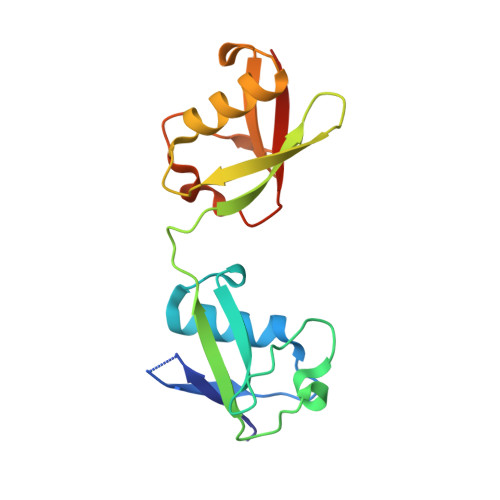
Structure of interferon-stimulated gene product 15 (ISG15) from the bat species Myotis davidii and the impact of interdomain ISG15 interactions on viral protein engagement.
Langley, C., Goodwin, O., Dzimianski, J.V., Daczkowski, C.M., Pegan, S.D.(2019) Acta Crystallogr D Struct Biol 75: 21-31
- PubMed: 30644842
- DOI: https://doi.org/10.1107/S2059798318015322
- Primary Citation of Related Structures:
6MDH - PubMed Abstract:
Bats have long been observed to be the hosts and the origin of numerous human diseases. Bats, like all mammals, rely on a number of innate immune mechanisms to combat invading pathogens, including the interferon type I, II and III responses. Ubiquitin-like interferon-stimulated gene product 15 (ISG15) is a key modulator of these interferon responses. Within these pathways, ISG15 can serve to stabilize host proteins modulating innate immune responses and act as a cytokine. Post-translational modifications of viral proteins introduced by ISG15 have also been observed to directly affect the function of numerous viral proteins. Unlike ubiquitin, which is virtually identical across all animals, comparison of ISG15s across species reveals that they are relatively divergent, with sequence identity dropping to as low as ∼58% among mammals. In addition to serving as an obstacle to the zoonotic transmission of influenza, these ISG15 species-species differences have also long been shown to have an impact on the function of viral deISGylases. Recently, the structure of the first nonhuman ISG15, originating from mouse, suggested that the structures of human ISG15 may not be reflective of other species. Here, the structure of ISG15 from the bat species Myotis davidii solved to 1.37 Å resolution is reported. Comparison of this ISG15 structure with those from human and mouse not only underscores the structural impact of ISG15 species-species differences, but also highlights a conserved hydrophobic motif formed between the two domains of ISG15. Using the papain-like deISGylase from Severe acute respiratory syndrome coronavirus as a probe, the biochemical importance of this motif in ISG15-protein engagements was illuminated.
Organizational Affiliation:
Pharmaceutical and Biomedical Sciences, University of Georgia, 240 West Green Street, Athens, GA 30602, USA.