6M35

Crystal structure of sulfur oxygenase reductase from Sulfurisphaera tokodaii


Experimental Data Snapshot

  • Method: X-RAY DIFFRACTION
  • Resolution: 1.73 Å
  • R-Value Free: 0.179 
  • R-Value Work: 0.162 
  • R-Value Observed: 0.163 

wwPDB Validation   3D Report Full Report


This is version 1.2 of the entry. See complete history


Literature

Crystallographic and cryogenic electron microscopic structures and enzymatic characterization of sulfur oxygenase reductase fromSulfurisphaera tokodaii.

Sato, Y.Yabuki, T.Adachi, N.Moriya, T.Arakawa, T.Kawasaki, M.Yamada, C.Senda, T.Fushinobu, S.Wakagi, T.

(2020) J Struct Biol X 4: 100030-100030

  • DOI: https://doi.org/10.1016/j.yjsbx.2020.100030
  • Primary Citation of Related Structures:  
    6M35, 6M3X

  • PubMed Abstract: 

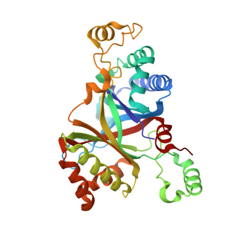
    Sulfur oxygenase reductases (SORs) are present in thermophilic and mesophilic archaea and bacteria, and catalyze oxygen-dependent oxygenation and disproportionation of elemental sulfur. SOR has a hollow, spherical homo-24-mer structure and reactions take place at active sites inside the chamber. The crystal structures of SORs from Acidianus species have been reported. However, the states of the active site components (mononuclear iron and cysteines) and the entry and exit paths of the substrate and products are still in dispute. Here, we report the biochemical and structural characterizations of SORs from the thermoacidophilic archaeon Sulfurisphaera tokodaii (StSOR) and present high-resolution structures determined by X-ray crystallography and cryogenic electron microscopy (cryo-EM). The crystal structure of StSOR was determined at 1.73 Å resolution. At the catalytic center, iron is ligated to His86, His90, Glu114, and two water molecules. Three conserved cysteines in the cavity are located 9.5-13 Å from the iron and were observed as free thiol forms. A mutational analysis indicated that the iron and one of the cysteines (Cys31) were essential for both activities. The cryo-EM structure was determined at 2.24 Å resolution using an instrument operating at 200 kV. The two structures determined by different methodologies showed similar main chain traces, but the maps exhibited different features at catalytically important components. A possible role of StSOR in the sulfur metabolism of S. tokodaii (an obligate aerobe) is discussed based on this study. Given the high resolution achieved in this study, StSOR was shown to be a good benchmark sample for cryo-EM.


  • Organizational Affiliation

    Department of Biotechnology, The University of Tokyo, 1-1-1 Yayoi, Bunkyo-ku, Tokyo 113-8657, Japan.


Macromolecules
Find similar proteins by:  (by identity cutoff)  |  3D Structure
Entity ID: 1
MoleculeChains Sequence LengthOrganismDetailsImage
Sulfur oxygenase/reductase
A, B, C, D, E
A, B, C, D, E, F, G, H
311Sulfurisphaera tokodaii str. 7Mutation(s): 0 
Gene Names: sorST1127STK_11270
EC: 1.13.11.55
UniProt
Find proteins for Q972K4 (Sulfurisphaera tokodaii (strain DSM 16993 / JCM 10545 / NBRC 100140 / 7))
Explore Q972K4 
Go to UniProtKB:  Q972K4
Entity Groups  
Sequence Clusters30% Identity50% Identity70% Identity90% Identity95% Identity100% Identity
UniProt GroupQ972K4
Sequence Annotations
Expand
  • Reference Sequence
Small Molecules
Ligands 3 Unique
IDChains Name / Formula / InChI Key2D Diagram3D Interactions
SO4
Query on SO4

Download Ideal Coordinates CCD File 
AA [auth F]
DA [auth G]
GA [auth H]
N [auth B]
Q [auth C]
AA [auth F],
DA [auth G],
GA [auth H],
N [auth B],
Q [auth C],
T [auth D],
W [auth E]
SULFATE ION
O4 S
QAOWNCQODCNURD-UHFFFAOYSA-L
GOL
Query on GOL

Download Ideal Coordinates CCD File 
CA [auth G]
FA [auth H]
J [auth A]
K [auth A]
M [auth B]
CA [auth G],
FA [auth H],
J [auth A],
K [auth A],
M [auth B],
P [auth C],
S [auth D],
V [auth E],
Y [auth F],
Z [auth F]
GLYCEROL
C3 H8 O3
PEDCQBHIVMGVHV-UHFFFAOYSA-N
FE (Subject of Investigation/LOI)
Query on FE

Download Ideal Coordinates CCD File 
BA [auth G]
EA [auth H]
I [auth A]
L [auth B]
O [auth C]
BA [auth G],
EA [auth H],
I [auth A],
L [auth B],
O [auth C],
R [auth D],
U [auth E],
X [auth F]
FE (III) ION
Fe
VTLYFUHAOXGGBS-UHFFFAOYSA-N
Experimental Data & Validation

Experimental Data

  • Method: X-RAY DIFFRACTION
  • Resolution: 1.73 Å
  • R-Value Free: 0.179 
  • R-Value Work: 0.162 
  • R-Value Observed: 0.163 
  • Space Group: I 2 3
Unit Cell:
Length ( Å )Angle ( ˚ )
a = 299.806α = 90
b = 299.806β = 90
c = 299.806γ = 90
Software Package:
Software NamePurpose
REFMACrefinement
PDB_EXTRACTdata extraction
XDSdata reduction
Aimlessdata scaling
MOLREPphasing

Structure Validation

View Full Validation Report


Entry History & Funding Information

Deposition Data


Funding OrganizationLocationGrant Number
Japan Society for the Promotion of Science (JSPS)Japan24580136
Japan Agency for Medical Research and Development (AMED)JapanJP20am0101071

Revision History  (Full details and data files)

  • Version 1.0: 2020-07-15
    Type: Initial release
  • Version 1.1: 2020-08-26
    Changes: Database references, Derived calculations
  • Version 1.2: 2023-11-29
    Changes: Data collection, Database references, Refinement description