Crystal Structure-Based Exploration of Arginine-Containing Peptide Binding in the ADP-Ribosyltransferase Domain of the Type III Effector XopAI Protein.
Liu, J.H., Yang, J.Y., Hsu, D.W., Lai, Y.H., Li, Y.P., Tsai, Y.R., Hou, M.H.(2019) Int J Mol Sci 20
- PubMed: 31615004
- DOI: https://doi.org/10.3390/ijms20205085
- Primary Citation of Related Structures:
6K93, 6K94, 6KLY - PubMed Abstract:
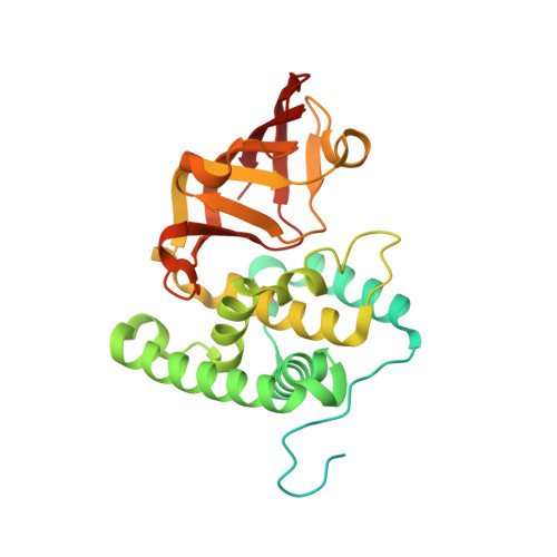
Plant pathogens secrete proteins called effectors into the cells of their host to modulate the host immune response against colonization. Effectors can either modify or arrest host target proteins to sabotage the signaling pathway, and therefore are considered potential drug targets for crop disease control. In earlier research, the Xanthomonas type III effector XopAI was predicted to be a member of the arginine-specific mono-ADP-ribosyltransferase family. However, the crystal structure of XopAI revealed an altered active site that is unsuitable to bind the cofactor NAD+, but with the capability to capture an arginine-containing peptide from XopAI itself. The arginine peptide consists of residues 60 through 69 of XopAI, and residue 62 (R62) is key to determining the protein-peptide interaction. The crystal structure and the molecular dynamics simulation results indicate that specific arginine recognition is mediated by hydrogen bonds provided by the backbone oxygen atoms from residues W154, T155, and T156, and a salt bridge provided by the E265 sidechain. In addition, a protruding loop of XopAI adopts dynamic conformations in response to arginine peptide binding and is probably involved in target protein recognition. These data suggest that XopAI binds to its target protein by the peptide-binding ability, and therefore, it promotes disease progression. Our findings reveal an unexpected and intriguing function of XopAI and pave the way for further investigation on the role of XopAI in pathogen invasion.
Organizational Affiliation:
Institute of Genomics and Bioinformatics, National Chung Hsing University (NCHU), Taichung 40227, Taiwan. jhliu@nchu.edu.tw.