6KC4

Crystal structure of human Fer SH2 domain bound to a phosphopeptide (DEpYENVD)


Experimental Data Snapshot

  • Method: X-RAY DIFFRACTION
  • Resolution: 1.37 Å
  • R-Value Free: 0.185 
  • R-Value Work: 0.165 
  • R-Value Observed: 0.166 

wwPDB Validation   3D Report Full Report


This is version 1.3 of the entry. See complete history


Literature

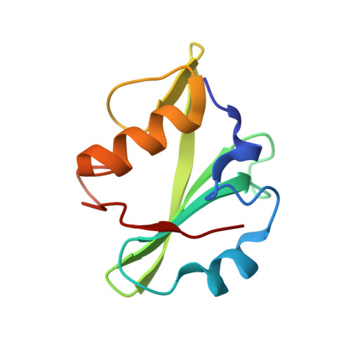
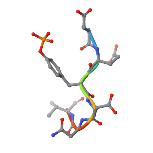
High-resolution structural analysis shows how different crystallographic environments can induce alternative modes of binding of a phosphotyrosine peptide to the SH2 domain of Fer tyrosine kinase.

Matsuura, Y.

(2019) Protein Sci 28: 2011-2019

  • DOI: https://doi.org/10.1002/pro.3713
  • Primary Citation of Related Structures:  
    6KC4

  • PubMed Abstract: 

    Fes and Fes-related (Fer) protein tyrosine kinases (PTKs) comprise a subfamily of nonreceptor tyrosine kinases characterized by a unique multidomain structure composed of an N-terminal Fer/CIP4 homology-Bin/Amphiphysin/Rvs (F-BAR) domain, a central Src homology 2 (SH2) domain, and a C-terminal PTK domain. Fer is ubiquitously expressed, and upregulation of Fer has been implicated in various human cancers. The PTK activity of Fes has been shown to be positively regulated by the binding of phosphotyrosine-containing ligands to the SH2 domain. Here, the X-ray crystal structure of human Fer SH2 domain bound to a phosphopeptide that has D-E-pY-E-N-V-D sequence is reported at 1.37 å resolution. The asymmetric unit (ASU) contains six Fer-phosphopeptide complexes, and the structure reveals three distinct binding modes for the same phosphopeptide. At four out of the six binding sites in the ASU, the phosphopeptide binds to Fer SH2 domain in a type I β-turn conformation, and this could be the optimal binding mode of this phosphopeptide. At the other two binding sites in the ASU, it appears that spatial proximity of neighboring SH2 domains in the crystal induces alternative modes of binding of this phosphopeptide.


  • Organizational Affiliation

    Division of Biological Science, Graduate School of Science, Nagoya University, Nagoya, Japan.


Macromolecules
Find similar proteins by:  (by identity cutoff)  |  3D Structure
Entity ID: 1
MoleculeChains Sequence LengthOrganismDetailsImage
Tyrosine-protein kinase Fer
A, C, E, G, I
A, C, E, G, I, K
102Homo sapiensMutation(s): 0 
Gene Names: FERTYK3
EC: 2.7.10.2
UniProt & NIH Common Fund Data Resources
Find proteins for P16591 (Homo sapiens)
Explore P16591 
Go to UniProtKB:  P16591
PHAROS:  P16591
GTEx:  ENSG00000151422 
Entity Groups  
Sequence Clusters30% Identity50% Identity70% Identity90% Identity95% Identity100% Identity
UniProt GroupP16591
Sequence Annotations
Expand
  • Reference Sequence

Find similar proteins by:  Sequence   |   3D Structure  

Entity ID: 2
MoleculeChains Sequence LengthOrganismDetailsImage
phosphopeptide (EDpYENVD)
B, D, F, H, J
B, D, F, H, J, L
7synthetic constructMutation(s): 0 
Sequence Annotations
Expand
  • Reference Sequence
Small Molecules
Modified Residues  1 Unique
IDChains TypeFormula2D DiagramParent
PTR
Query on PTR
B, D, F, H, J
B, D, F, H, J, L
L-PEPTIDE LINKINGC9 H12 N O6 PTYR
Experimental Data & Validation

Experimental Data

  • Method: X-RAY DIFFRACTION
  • Resolution: 1.37 Å
  • R-Value Free: 0.185 
  • R-Value Work: 0.165 
  • R-Value Observed: 0.166 
  • Space Group: P 1 21 1
Unit Cell:
Length ( Å )Angle ( ˚ )
a = 34.7α = 90
b = 186.661β = 96.96
c = 50.58γ = 90
Software Package:
Software NamePurpose
Aimlessdata scaling
MOLREPphasing
PHENIXrefinement
PDB_EXTRACTdata extraction
MOSFLMdata reduction

Structure Validation

View Full Validation Report


Entry History & Funding Information

Deposition Data

  • Released Date: 2019-09-04 
  • Deposition Author(s): Matsuura, Y.

Funding OrganizationLocationGrant Number
Japan Society for the Promotion of ScienceJapan16K07268

Revision History  (Full details and data files)

  • Version 1.0: 2019-09-04
    Type: Initial release
  • Version 1.1: 2019-09-11
    Changes: Data collection, Database references
  • Version 1.2: 2019-10-30
    Changes: Data collection, Database references
  • Version 1.3: 2023-11-22
    Changes: Data collection, Database references, Refinement description