6JOD

Angiotensin II type 2 receptor with ligand


Experimental Data Snapshot

  • Method: X-RAY DIFFRACTION
  • Resolution: 3.20 Å
  • R-Value Free: 0.287 
  • R-Value Work: 0.235 
  • R-Value Observed: 0.237 

wwPDB Validation   3D Report Full Report


This is version 1.2 of the entry. See complete history


Literature

The Crystal Structure of Angiotensin II Type 2 Receptor with Endogenous Peptide Hormone.

Asada, H.Inoue, A.Ngako Kadji, F.M.Hirata, K.Shiimura, Y.Im, D.Shimamura, T.Nomura, N.Iwanari, H.Hamakubo, T.Kusano-Arai, O.Hisano, H.Uemura, T.Suno, C.Aoki, J.Iwata, S.

(2020) Structure 28: 418

  • DOI: https://doi.org/10.1016/j.str.2019.12.003
  • Primary Citation of Related Structures:  
    6JOD

  • PubMed Abstract: 

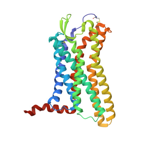
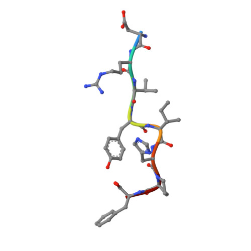
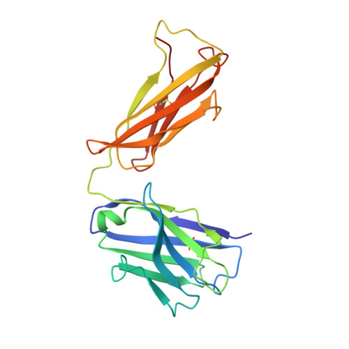
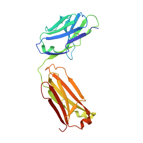
    Angiotensin II (AngII) is a peptide hormone that plays a key role in regulating blood pressure, and its interactions with the G protein-coupled receptors, AngII type-1 receptor (AT 1 R) and AngII type-2 receptor (AT 2 R), are central to its mechanism of action. We solved the crystal structure of human AT 2 R bound to AngII and its specific antibody at 3.2-Å resolution. AngII (full agonist) and [Sar 1 , Ile 8 ]-AngII (partial agonist) interact with AT 2 R in a similar fashion, except at the bottom of the AT 2 R ligand-binding pocket. In particular, the residues including Met128 3.36 , which constitute the deep end of the cavity, play important roles in angiotensin receptor (ATR) activation upon AngII binding. These differences that occur upon endogenous ligand binding may contribute to a structural change in AT 2 R, leading to normalization of the non-canonical coordination of helix 8. Our results will inform the design of more effective ligands for ATRs.


  • Organizational Affiliation

    Department of Cell Biology, Graduate School of Medicine, Kyoto University, Kyoto 606-8501, Japan. Electronic address: asada.hidetsugu.4s@kyoto-u.ac.jp.


Macromolecules
Find similar proteins by:  (by identity cutoff)  |  3D Structure
Entity ID: 1
MoleculeChains Sequence LengthOrganismDetailsImage
Type-2 angiotensin II receptor312Homo sapiensMutation(s): 1 
Gene Names: AGTR2
Membrane Entity: Yes 
UniProt & NIH Common Fund Data Resources
Find proteins for P50052 (Homo sapiens)
Explore P50052 
Go to UniProtKB:  P50052
PHAROS:  P50052
GTEx:  ENSG00000180772 
Entity Groups  
Sequence Clusters30% Identity50% Identity70% Identity90% Identity95% Identity100% Identity
UniProt GroupP50052
Sequence Annotations
Expand
  • Reference Sequence

Find similar proteins by:  Sequence   |   3D Structure  

Entity ID: 2
MoleculeChains Sequence LengthOrganismDetailsImage
Angiotensin II8Homo sapiensMutation(s): 0 
Membrane Entity: Yes 
UniProt & NIH Common Fund Data Resources
Find proteins for P01019 (Homo sapiens)
Explore P01019 
Go to UniProtKB:  P01019
PHAROS:  P01019
GTEx:  ENSG00000135744 
Entity Groups  
UniProt GroupP01019
Sequence Annotations
Expand
  • Reference Sequence
Find similar proteins by:  (by identity cutoff)  |  3D Structure
Entity ID: 3
MoleculeChains Sequence LengthOrganismDetailsImage
Soluble cytochrome b562,Soluble cytochrome b56286Escherichia coliMutation(s): 4 
Gene Names: cybC
Membrane Entity: Yes 
UniProt
Find proteins for P0ABE7 (Escherichia coli)
Explore P0ABE7 
Go to UniProtKB:  P0ABE7
Entity Groups  
Sequence Clusters30% Identity50% Identity70% Identity90% Identity95% Identity100% Identity
UniProt GroupP0ABE7
Sequence Annotations
Expand
  • Reference Sequence
Find similar proteins by:  (by identity cutoff)  |  3D Structure
Entity ID: 4
MoleculeChains Sequence LengthOrganismDetailsImage
Heavy chain of 4A03FabD [auth H]220Mus musculusMutation(s): 0 
Entity Groups  
Sequence Clusters30% Identity50% Identity70% Identity90% Identity95% Identity100% Identity
Sequence Annotations
Expand
  • Reference Sequence
Find similar proteins by:  (by identity cutoff)  |  3D Structure
Entity ID: 5
MoleculeChains Sequence LengthOrganismDetailsImage
Light chain of 4A03FabE [auth L]212Mus musculusMutation(s): 0 
Entity Groups  
Sequence Clusters30% Identity50% Identity70% Identity90% Identity95% Identity100% Identity
Sequence Annotations
Expand
  • Reference Sequence
Experimental Data & Validation

Experimental Data

  • Method: X-RAY DIFFRACTION
  • Resolution: 3.20 Å
  • R-Value Free: 0.287 
  • R-Value Work: 0.235 
  • R-Value Observed: 0.237 
  • Space Group: C 2 2 21
Unit Cell:
Length ( Å )Angle ( ˚ )
a = 102.55α = 90
b = 426.43β = 90
c = 52.7γ = 90
Software Package:
Software NamePurpose
PHENIXrefinement
XSCALEdata scaling
PDB_EXTRACTdata extraction
XDSdata reduction
MOLREPphasing

Structure Validation

View Full Validation Report


Entry History & Funding Information

Deposition Data


Funding OrganizationLocationGrant Number
Japan Agency for Medical Research and Development (AMED)JapanJP18am0101070

Revision History  (Full details and data files)

  • Version 1.0: 2020-01-15
    Type: Initial release
  • Version 1.1: 2020-04-22
    Changes: Database references
  • Version 1.2: 2023-11-22
    Changes: Data collection, Database references, Refinement description