6JJJ

Trimeric structure of Kupffer cell C-type lectin receptor Clec4f


Experimental Data Snapshot

  • Method: X-RAY DIFFRACTION
  • Resolution: 2.79 Å
  • R-Value Free: 0.292 
  • R-Value Work: 0.250 
  • R-Value Observed: 0.254 

wwPDB Validation   3D Report Full Report


This is version 1.2 of the entry. See complete history


Literature

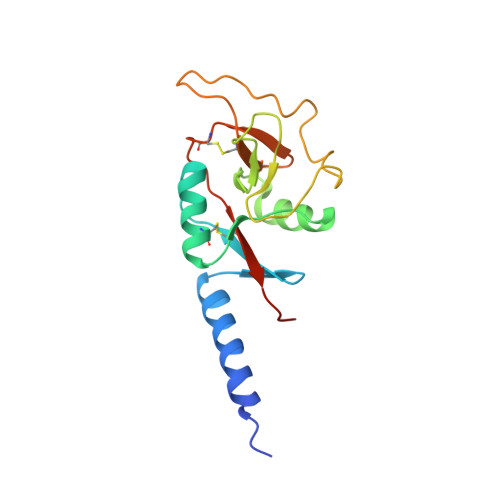
Trimeric structure of the mouse Kupffer cell C-type lectin receptor Clec4f.

Ouyang, Z.Felix, J.Zhou, J.Pei, Y.Ma, B.Hwang, P.M.Lemieux, M.J.Gutsche, I.Zheng, F.Wen, Y.

(2020) FEBS Lett 594: 189-198

  • DOI: https://doi.org/10.1002/1873-3468.13565
  • Primary Citation of Related Structures:  
    6JJJ

  • PubMed Abstract: 

    The C-type lectin receptor Clec4f has been identified as a specific surface marker for Kupffer cells, although its ortholog is absent in humans and its biological function remains elusive. Here, we report the crystal structure of a truncated mouse trimeric Clec4f. The orientation between the carbohydrate-recognition domain of Clec4f and its neck region differs from other C-type lectins, resulting in an observed distance of 45 Å between the glycan-binding sites within the Clec4f trimer. Interestingly, the trimeric coiled-coil interface within its heptad neck region contains multiple polyglutamine interactions instead of the predominantly hydrophobic leucine zipper found in other C-type lectin receptors. The Clec4f trimeric structure displays unique features regarding its assembly and ligand recognition, shedding light on the evolution and diversity of the C-type lectin family.


  • Organizational Affiliation

    The Key Laboratory of Biomedical Information Engineering of Ministry of Education, School of Life Science and Technology, Xi'an Jiaotong University, China.


Macromolecules
Find similar proteins by:  (by identity cutoff)  |  3D Structure
Entity ID: 1
MoleculeChains Sequence LengthOrganismDetailsImage
C-type lectin domain family 4 member F
A, B, C, D, E
A, B, C, D, E, F
160Mus musculusMutation(s): 0 
Gene Names: Clec4fClecsf13Kclr
UniProt
Find proteins for P70194 (Mus musculus)
Explore P70194 
Go to UniProtKB:  P70194
Entity Groups  
Sequence Clusters30% Identity50% Identity70% Identity90% Identity95% Identity100% Identity
UniProt GroupP70194
Sequence Annotations
Expand
  • Reference Sequence
Small Molecules
Ligands 1 Unique
IDChains Name / Formula / InChI Key2D Diagram3D Interactions
CA
Query on CA

Download Ideal Coordinates CCD File 
G [auth A]
H [auth A]
I [auth B]
J [auth B]
K [auth C]
G [auth A],
H [auth A],
I [auth B],
J [auth B],
K [auth C],
L [auth C],
M [auth D],
N [auth D],
O [auth E],
P [auth E],
Q [auth F],
R [auth F]
CALCIUM ION
Ca
BHPQYMZQTOCNFJ-UHFFFAOYSA-N
Experimental Data & Validation

Experimental Data

  • Method: X-RAY DIFFRACTION
  • Resolution: 2.79 Å
  • R-Value Free: 0.292 
  • R-Value Work: 0.250 
  • R-Value Observed: 0.254 
  • Space Group: P 1
Unit Cell:
Length ( Å )Angle ( ˚ )
a = 75.15α = 90.01
b = 75.19β = 90.04
c = 61.2γ = 120.06
Software Package:
Software NamePurpose
PHENIXrefinement
XDSdata reduction
XDSdata scaling
MOLREPphasing

Structure Validation

View Full Validation Report


Entry History & Funding Information

Deposition Data


Funding OrganizationLocationGrant Number
National Natural Science Foundation of ChinaChinaNO.31870132 and NO.81741088

Revision History  (Full details and data files)

  • Version 1.0: 2019-08-14
    Type: Initial release
  • Version 1.1: 2020-01-29
    Changes: Database references
  • Version 1.2: 2023-11-22
    Changes: Data collection, Database references, Derived calculations, Refinement description