6J3Q

Capsid structure of a freshwater cyanophage Siphoviridae Mic1


Experimental Data Snapshot

  • Method: ELECTRON MICROSCOPY
  • Resolution: 3.53 Å
  • Aggregation State: PARTICLE 
  • Reconstruction Method: SINGLE PARTICLE 

wwPDB Validation   3D Report Full Report


This is version 1.3 of the entry. See complete history


Literature

Capsid Structure of a Freshwater Cyanophage Siphoviridae Mic1.

Jin, H.Jiang, Y.L.Yang, F.Zhang, J.T.Li, W.F.Zhou, K.Ju, J.Chen, Y.Zhou, C.Z.

(2019) Structure 27: 1508

  • DOI: https://doi.org/10.1016/j.str.2019.07.003
  • Primary Citation of Related Structures:  
    6J3Q

  • PubMed Abstract: 

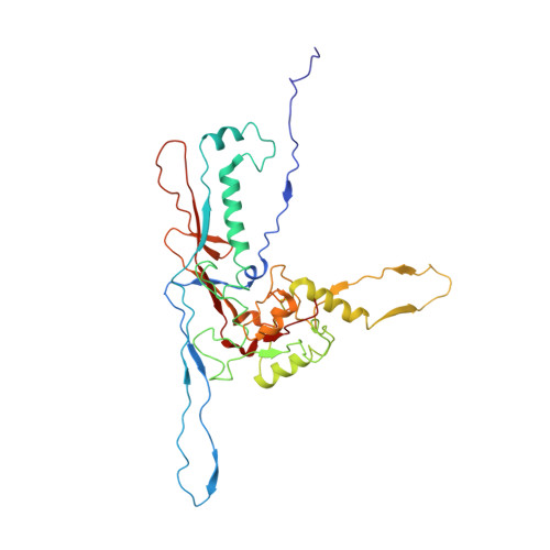
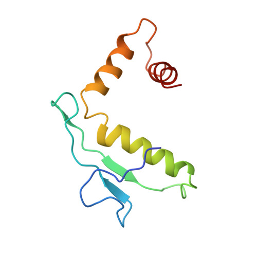
    Cyanobacteria are the most abundant photosynthetic microorganisms, the global distribution of which is mainly regulated by the corresponding cyanophages. A systematic screening of water samples in the Lake Chaohu enabled us to isolate a freshwater siphocyanophage that infects Microcystis wesenbergii, thus termed Mic1. Using cryoelectron microscopy, we solved the 3.5-Å structure of Mic1 capsid. The major capsid protein gp40 of an HK97-like fold forms two types of capsomers, hexons and pentons. The capsomers interact with each other via the interweaved N-terminal arms of gp40 in addition to a tail-in-mouth joint along the three-fold symmetric axis, resulting in the assembly of capsid in a mortise-and-tenon pattern. The novel-fold cement protein gp47 sticks at the two-fold symmetric axis and further fixes the capsid. These findings provide structural insights into the assembly of cyanophages, and set up a platform to explore the mechanism of specific interactions and co-evolution with cyanobacteria.


  • Organizational Affiliation

    Hefei National Laboratory for Physical Sciences at the Microscale and School of Life Sciences, University of Science and Technology of China, Hefei, Anhui 230026, China.


Macromolecules
Find similar proteins by:  (by identity cutoff)  |  3D Structure
Entity ID: 1
MoleculeChains Sequence LengthOrganismDetailsImage
major capsid protein348Microcystis phage Mic1Mutation(s): 0 
UniProt
Find proteins for A0A4Y5TR23 (Microcystis phage Mic1)
Explore A0A4Y5TR23 
Go to UniProtKB:  A0A4Y5TR23
Entity Groups  
Sequence Clusters30% Identity50% Identity70% Identity90% Identity95% Identity100% Identity
UniProt GroupA0A4Y5TR23
Sequence Annotations
Expand
  • Reference Sequence
Find similar proteins by:  (by identity cutoff)  |  3D Structure
Entity ID: 2
MoleculeChains Sequence LengthOrganismDetailsImage
cement protein99Microcystis phage Mic1Mutation(s): 0 
UniProt
Find proteins for A0A4Y5TPY8 (Microcystis phage Mic1)
Explore A0A4Y5TPY8 
Go to UniProtKB:  A0A4Y5TPY8
Entity Groups  
Sequence Clusters30% Identity50% Identity70% Identity90% Identity95% Identity100% Identity
UniProt GroupA0A4Y5TPY8
Sequence Annotations
Expand
  • Reference Sequence
Experimental Data & Validation

Experimental Data

  • Method: ELECTRON MICROSCOPY
  • Resolution: 3.53 Å
  • Aggregation State: PARTICLE 
  • Reconstruction Method: SINGLE PARTICLE 
EM Software:
TaskSoftware PackageVersion
MODEL REFINEMENTPHENIX
RECONSTRUCTIONRELION2.0

Structure Validation

View Full Validation Report


Entry History & Funding Information

Deposition Data


Funding OrganizationLocationGrant Number
National Natural Science Foundation of ChinaChina31630001
National Natural Science Foundation of ChinaChina31621002
Ministry of Science and Technology (China)China2016YFA0400900
Chinese Academy of SciencesChina--

Revision History  (Full details and data files)

  • Version 1.0: 2019-10-02
    Type: Initial release
  • Version 1.1: 2019-10-16
    Changes: Data collection, Database references
  • Version 1.2: 2019-11-27
    Changes: Source and taxonomy
  • Version 1.3: 2024-03-27
    Changes: Data collection, Database references, Derived calculations