


FBIs complex structure of peptide deformylase from Xanthomonas oryzae pv. oryzae
Lee, I.H., Kang, L.W.To be published.
Experimental Data Snapshot
Entity ID: 1 | |||||
|---|---|---|---|---|---|
| Molecule | Chains | Sequence Length | Organism | Details | Image |
| Peptide deformylase | 167 | Xanthomonas oryzae pv. oryzae KACC 10331 | Mutation(s): 0 Gene Names: def, XOO1075 EC: 3.5.1.88 | 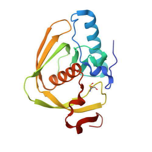 | |
UniProt | |||||
Find proteins for Q5H3Z2 (Xanthomonas oryzae pv. oryzae (strain KACC10331 / KXO85)) Explore Q5H3Z2 Go to UniProtKB: Q5H3Z2 | |||||
Entity Groups | |||||
| Sequence Clusters | 30% Identity50% Identity70% Identity90% Identity95% Identity100% Identity | ||||
| UniProt Group | Q5H3Z2 | ||||
Sequence AnnotationsExpand | |||||
| |||||
| Ligands 2 Unique | |||||
|---|---|---|---|---|---|
| ID | Chains | Name / Formula / InChI Key | 2D Diagram | 3D Interactions | |
| LHY (Subject of Investigation/LOI) Query on LHY | E [auth A] | L-[(N-HYDROXYAMINO)CARBONYL]PHENYLALANINE C10 H12 N2 O4 IOFPEOPOAMOMBE-QMMMGPOBSA-N |  | ||
| CD Query on CD | B [auth A], C [auth A], D [auth A] | CADMIUM ION Cd WLZRMCYVCSSEQC-UHFFFAOYSA-N |  | ||
| Modified Residues 1 Unique | |||||
|---|---|---|---|---|---|
| ID | Chains | Type | Formula | 2D Diagram | Parent |
| CSD Query on CSD | A | L-PEPTIDE LINKING | C3 H7 N O4 S |  | CYS |
| Length ( Å ) | Angle ( ˚ ) |
|---|---|
| a = 58.531 | α = 90 |
| b = 58.531 | β = 90 |
| c = 266.212 | γ = 120 |
| Software Name | Purpose |
|---|---|
| HKL-2000 | data scaling |
| REFMAC | refinement |
| HKL-2000 | data collection |
| HKL-2000 | data reduction |
RCSB PDB (citation) is hosted by
RCSB PDB is a member of the

