


Crystal structure of an uncharacterized protein
Li, B.To be published.
Experimental Data Snapshot
Entity ID: 1 | |||||
|---|---|---|---|---|---|
| Molecule | Chains | Sequence Length | Organism | Details | Image |
| UPF0061 protein YdiU | 482 | Escherichia coli | Mutation(s): 0 Gene Names: ydiU, BMT91_02235, NCTC9073_05692, PGD_01529, SAMEA3472078_00997, SAMEA3484419_01283, SAMEA3752562_01353, SAMEA3753069_03695, SAMEA3753077_01245 | 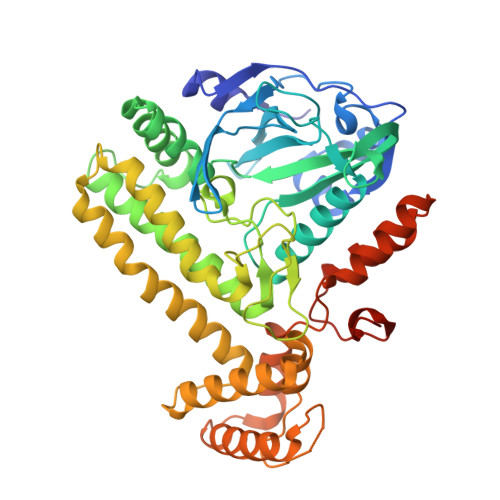 | |
UniProt | |||||
Find proteins for P77649 (Escherichia coli (strain K12)) Explore P77649 Go to UniProtKB: P77649 | |||||
Entity Groups | |||||
| Sequence Clusters | 30% Identity50% Identity70% Identity90% Identity95% Identity100% Identity | ||||
| UniProt Group | P77649 | ||||
Sequence AnnotationsExpand | |||||
| |||||
| Ligands 3 Unique | |||||
|---|---|---|---|---|---|
| ID | Chains | Name / Formula / InChI Key | 2D Diagram | 3D Interactions | |
| AMP Query on AMP | C [auth A] | ADENOSINE MONOPHOSPHATE C10 H14 N5 O7 P UDMBCSSLTHHNCD-KQYNXXCUSA-N |  | ||
| 2PN Query on 2PN | B [auth A] | IMIDODIPHOSPHORIC ACID H5 N O6 P2 GNGSOPFGGKKDQP-UHFFFAOYSA-N |  | ||
| MG Query on MG | D [auth A], E [auth A] | MAGNESIUM ION Mg JLVVSXFLKOJNIY-UHFFFAOYSA-N |  | ||
| Length ( Å ) | Angle ( ˚ ) |
|---|---|
| a = 71.424 | α = 90 |
| b = 71.424 | β = 90 |
| c = 363.446 | γ = 120 |
| Software Name | Purpose |
|---|---|
| PHENIX | refinement |
| HKL-2000 | data reduction |
| HKL-2000 | data scaling |
| PHENIX | phasing |
RCSB PDB (citation) is hosted by
RCSB PDB is a member of the

