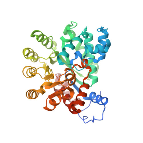
Crystal structure of Plasmodium falciparum adenosine deaminase reveals a novel binding pocket for inosine.
Jaruwat, A., Riangrungroj, P., Ubonprasert, S., Sae-Ueng, U., Kuaprasert, B., Yuthavong, Y., Leartsakulpanich, U., Chitnumsub, P.(2019) Arch Biochem Biophys 667: 6-13
- PubMed: 31002765
- DOI: https://doi.org/10.1016/j.abb.2019.04.002
- Primary Citation of Related Structures:
6II7 - PubMed Abstract:
Plasmodium falciparum (Pf), a malarial pathogen, can only synthesize purine nucleotides employing a salvage pathway because it lacks de novo biosynthesis. Adenosine deaminase (ADA), one of the three purine salvage enzymes, catalyzes the irreversible hydrolytic deamination of adenosine to inosine, which is further converted to GMP and AMP for DNA/RNA production. In addition to adenosine conversion, Plasmodium ADA also catalyzes the conversion of 5'-methylthioadenosine, derived from polyamine biosynthesis, into 5'-methylthioinosine whereas the human enzyme is not capable of this function. Here we report the crystal structure of a surface engineered PfADA at a resolution of 2.48 Å, together with results on kinetic studies of PfADA wild-type and active site variants. The structure reveals a novel inosine binding pocket linked to a distinctive PfADA substructure (residues 172-179) derived from a non-conserved gating helix loop (172-188) in Plasmodium spp. and other ADA enzymes. Variants of PfADA and human (h) ADA active site amino acids were generated in order to study their role in catalysis, including PfADA- Phe136, -Thr174, -Asp176, and -Leu179, and hADA-Met155, equivalent to PfADA-Asp176. PfADA-Leu179His showed no effect on kinetic parameters. However, kinetic results of PfADA-Asp176Met/Ala mutants and hADA-Met155Asp/Ala showed that the mutation reduced adenosine and 5'-methylthioadenosine substrate affinity in PfADA and k cat in hADA, thereby reducing catalytic efficiency of the enzyme. Phe136Leu mutant showed increased K m (>10-fold) for both substrates whereas Thr174Ile/Ala only affected 5'-methylthioadenosine binding affinity. Together, the structure with the novel inosine binding pocket and the kinetic data provide insights for rational design of inhibitors against PfADA.
Organizational Affiliation:
National Center for Genetic Engineering and Biotechnology, 113 Thailand Science Park, Phahonyothin Road, Klong 1, Klong Luang, Pathumthani, 12120, Thailand.