6IF6

Structure of the periplasmic domain of SflA


Experimental Data Snapshot

  • Method: X-RAY DIFFRACTION
  • Resolution: 1.90 Å
  • R-Value Free: 0.221 
  • R-Value Work: 0.176 
  • R-Value Observed: 0.180 

wwPDB Validation   3D Report Full Report


This is version 1.3 of the entry. See complete history


Literature

Structure of the periplasmic domain of SflA involved in spatial regulation of the flagellar biogenesis of Vibrio reveals a TPR/SLR-like fold.

Sakuma, M.Nishikawa, S.Inaba, S.Nishigaki, T.Kojima, S.Homma, M.Imada, K.

(2019) J Biochem 166: 197-204

  • DOI: https://doi.org/10.1093/jb/mvz027
  • Primary Citation of Related Structures:  
    6IF6

  • PubMed Abstract: 

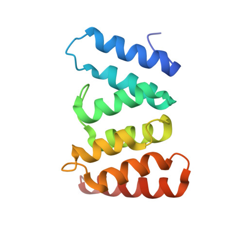
    Bacteria have evolved various types of flagellum, an organella for bacterial motility, to adapt to their habitat environments. The number and the spatial arrangement of the flagellum are precisely controlled to optimize performance of each type of the flagellar system. Vibrio alginolyticus has a single sheathed flagellum at the cell pole for swimming. SflA is a regulator protein to prevent peritrichous formation of the sheathed flagellum, and consists of an N-terminal periplasmic region, a transmembrane helix, and a C-terminal cytoplasmic region. Whereas the cytoplasmic region has been characterized to be essential for inhibition of the peritrichous growth, the role of the N-terminal region is still unclear. We here determined the structure of the N-terminal periplasmic region of SflA (SflAN) at 1.9-Å resolution. The core of SflAN forms a domain-swapped dimer with tetratricopeptide repeat (TPR)/Sel1-like repeat (SLR) motif, which is often found in the domains responsible for protein-protein interaction in various proteins. The structural similarity and the following mutational analysis based on the structure suggest that SflA binds to unknown partner protein by SflAN and the binding signal is important for the precise control of the SflA function.


  • Organizational Affiliation

    Radioisotope Research Center, Graduate School of Science, Nagoya University, Chikusa-Ku, Furocho, Nagoya, Japan.


Macromolecules
Find similar proteins by:  (by identity cutoff)  |  3D Structure
Entity ID: 1
MoleculeChains Sequence LengthOrganismDetailsImage
Protein SflA133Vibrio alginolyticusMutation(s): 0 
UniProt
Find proteins for A0A1W6TB41 (Vibrio alginolyticus)
Explore A0A1W6TB41 
Go to UniProtKB:  A0A1W6TB41
Entity Groups  
Sequence Clusters30% Identity50% Identity70% Identity90% Identity95% Identity100% Identity
UniProt GroupA0A1W6TB41
Sequence Annotations
Expand
  • Reference Sequence
Experimental Data & Validation

Experimental Data

  • Method: X-RAY DIFFRACTION
  • Resolution: 1.90 Å
  • R-Value Free: 0.221 
  • R-Value Work: 0.176 
  • R-Value Observed: 0.180 
  • Space Group: P 31 2 1
Unit Cell:
Length ( Å )Angle ( ˚ )
a = 66.9α = 90
b = 66.9β = 90
c = 59.8γ = 120
Software Package:
Software NamePurpose
PHENIXrefinement
MOSFLMdata reduction
Aimlessdata scaling
PHENIXphasing

Structure Validation

View Full Validation Report


Entry History & Funding Information

Deposition Data


Funding OrganizationLocationGrant Number
Japan Society for the Promotion of ScienceJapanJP15H02386
Japan Society for the Promotion of ScienceJapanJP16H04774

Revision History  (Full details and data files)

  • Version 1.0: 2019-05-01
    Type: Initial release
  • Version 1.1: 2019-05-15
    Changes: Data collection, Database references, Source and taxonomy, Structure summary
  • Version 1.2: 2019-08-14
    Changes: Data collection, Database references
  • Version 1.3: 2024-03-27
    Changes: Data collection, Database references