


6IBI
Copper binding protein from Laetisaria arvalis (LaX325)
- PDB DOI: https://doi.org/10.2210/pdb6IBI/pdb
- Classification: METAL BINDING PROTEIN
- Organism(s): Laetisaria arvalis
- Expression System: Komagataella pastoris
- Mutation(s): No
- Deposited: 2018-11-30 Released: 2019-11-13
- Funding Organization(s): Novo Nordisk Foundation, The Carslberg Foundation, European Communitys Seventh Framework Programme
Experimental Data Snapshot
- Method: X-RAY DIFFRACTION
- Resolution: 2.08 Å
- R-Value Free: 0.271
- R-Value Work: 0.244
- R-Value Observed: 0.245

This is version 1.4 of the entry. See complete history.

Macromolecules
Find similar proteins by:
(by identity cutoff) | 3D Structure
Entity ID: 1 | |||||
|---|---|---|---|---|---|
| Molecule | Chains | Sequence Length | Organism | Details | Image |
| Auxiliary activity CAZyme | 155 | Laetisaria arvalis | Mutation(s): 0 | 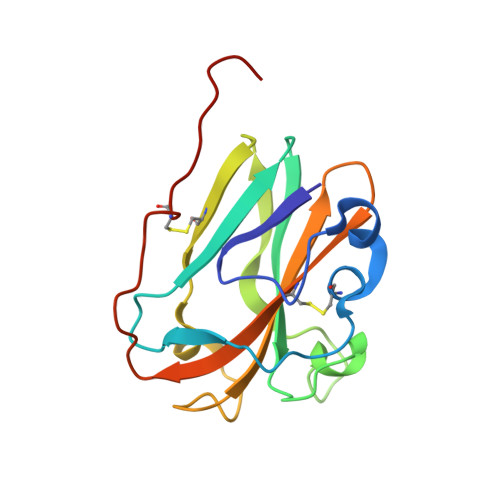 | |
UniProt | |||||
Find proteins for A0A4P9I8G4 (Waitea arvalis) Explore A0A4P9I8G4 Go to UniProtKB: A0A4P9I8G4 | |||||
Entity Groups | |||||
| Sequence Clusters | 30% Identity50% Identity70% Identity90% Identity95% Identity100% Identity | ||||
| UniProt Group | A0A4P9I8G4 | ||||
Sequence AnnotationsExpand | |||||
| |||||
Small Molecules
| Ligands 5 Unique | |||||
|---|---|---|---|---|---|
| ID | Chains | Name / Formula / InChI Key | 2D Diagram | 3D Interactions | |
| 1PE Query on 1PE | FA [auth C], L [auth A], MA [auth D], N [auth A], W [auth B] | PENTAETHYLENE GLYCOL C10 H22 O6 JLFNLZLINWHATN-UHFFFAOYSA-N |  | ||
| NAG Query on NAG | AA [auth C] BA [auth C] CA [auth C] DA [auth C] F [auth A] | 2-acetamido-2-deoxy-beta-D-glucopyranose C8 H15 N O6 OVRNDRQMDRJTHS-FMDGEEDCSA-N |  | ||
| PGE Query on PGE | M [auth A] | TRIETHYLENE GLYCOL C6 H14 O4 ZIBGPFATKBEMQZ-UHFFFAOYSA-N |  | ||
| IMD Query on IMD | EA [auth C], V [auth B] | IMIDAZOLE C3 H5 N2 RAXXELZNTBOGNW-UHFFFAOYSA-O |  | ||
| CU Query on CU | E [auth A], GA [auth D], O [auth B], X [auth C] | COPPER (II) ION Cu JPVYNHNXODAKFH-UHFFFAOYSA-N |  | ||
Experimental Data & Validation
Experimental Data
- Method: X-RAY DIFFRACTION
- Resolution: 2.08 Å
- R-Value Free: 0.271
- R-Value Work: 0.244
- R-Value Observed: 0.245
- Space Group: P 21 21 21
Unit Cell:
| Length ( Å ) | Angle ( ˚ ) |
|---|---|
| a = 84.599 | α = 90 |
| b = 84.635 | β = 90 |
| c = 127.319 | γ = 90 |
| Software Name | Purpose |
|---|---|
| REFMAC | refinement |
| MxCuBE | data collection |
| XDS | data reduction |
| XSCALE | data scaling |
| PHENIX | phasing |
| AutoSol | phasing |
| PHASER | phasing |
| Coot | model building |
Entry History & Funding Information
Deposition Data
- Released Date: 2019-11-13 Deposition Author(s): Frandsen, K.E.H., Tandrup, T., Labourel, A., Haon, M., Berrin, J.-G., Lo Leggio, L.
| Funding Organization | Location | Grant Number |
|---|---|---|
| Novo Nordisk Foundation | Denmark | NF17SA0027704 |
| The Carslberg Foundation | Denmark | CF16-0673 & CF17-0533 |
| European Communitys Seventh Framework Programme | France | grant agreement 609398 |
Revision History (Full details and data files)
- Version 1.0: 2019-11-13
Type: Initial release - Version 1.1: 2019-11-27
Changes: Derived calculations - Version 1.2: 2020-01-22
Changes: Database references - Version 1.3: 2020-03-04
Changes: Data collection, Database references - Version 1.4: 2020-07-29
Type: Remediation
Reason: Carbohydrate remediation
Changes: Data collection, Derived calculations, Structure summary
