Crystal structures of the Bacillus subtilis prophage lytic cassette proteins XepA and YomS.
Freitag-Pohl, S., Jasilionis, A., Hakansson, M., Svensson, L.A., Kovacic, R., Welin, M., Watzlawick, H., Wang, L., Altenbuchner, J., Plotka, M., Kaczorowska, A.K., Kaczorowski, T., Nordberg Karlsson, E., Al-Karadaghi, S., Walse, B., Aevarsson, A., Pohl, E.(2019) Acta Crystallogr D Struct Biol 75: 1028-1039
- PubMed: 31692476
- DOI: https://doi.org/10.1107/S2059798319013330
- Primary Citation of Related Structures:
6I56, 6I5O, 6IA5 - PubMed Abstract:
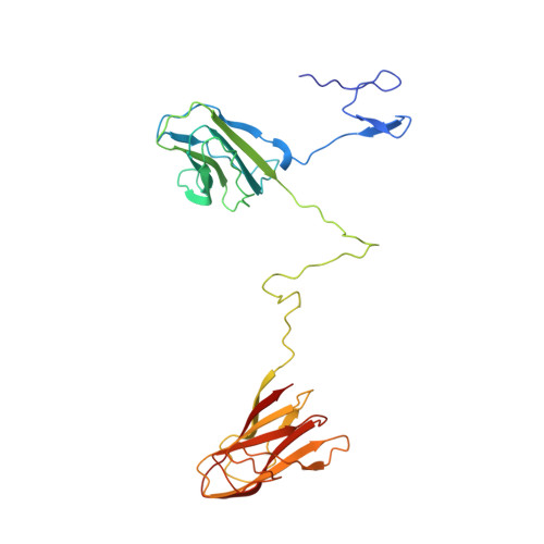
As part of the Virus-X Consortium that aims to identify and characterize novel proteins and enzymes from bacteriophages and archaeal viruses, the genes of the putative lytic proteins XepA from Bacillus subtilis prophage PBSX and YomS from prophage SPβ were cloned and the proteins were subsequently produced and functionally characterized. In order to elucidate the role and the molecular mechanism of XepA and YomS, the crystal structures of these proteins were solved at resolutions of 1.9 and 1.3 Å, respectively. XepA consists of two antiparallel β-sandwich domains connected by a 30-amino-acid linker region. A pentamer of this protein adopts a unique dumbbell-shaped architecture consisting of two discs and a central tunnel. YomS (12.9 kDa per monomer), which is less than half the size of XepA (30.3 kDa), shows homology to the C-terminal part of XepA and exhibits a similar pentameric disc arrangement. Each β-sandwich entity resembles the fold of typical cytoplasmic membrane-binding C2 domains. Only XepA exhibits distinct cytotoxic activity in vivo, suggesting that the N-terminal pentameric domain is essential for this biological activity. The biological and structural data presented here suggest that XepA disrupts the proton motive force of the cytoplasmatic membrane, thus supporting cell lysis.
Organizational Affiliation:
Department of Chemistry, Durham University, South Road, Durham DH1 3LE, England.