6I2K

Structure of EV71 complexed with its receptor SCARB2


Experimental Data Snapshot

  • Method: ELECTRON MICROSCOPY
  • Resolution: 3.40 Å
  • Aggregation State: PARTICLE 
  • Reconstruction Method: SINGLE PARTICLE 

wwPDB Validation   3D Report Full Report


This is version 2.0 of the entry. See complete history


Literature

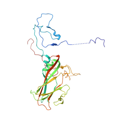
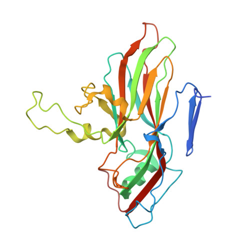
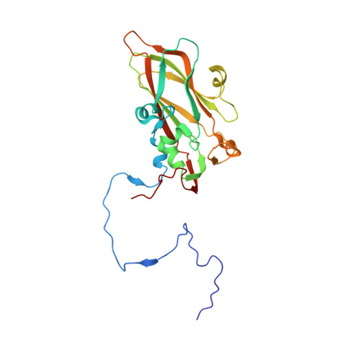
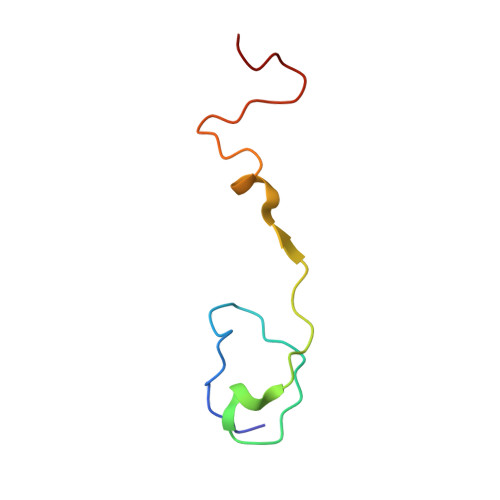
Unexpected mode of engagement between enterovirus 71 and its receptor SCARB2.

Zhou, D.Zhao, Y.Kotecha, A.Fry, E.E.Kelly, J.T.Wang, X.Rao, Z.Rowlands, D.J.Ren, J.Stuart, D.I.

(2019) Nat Microbiol 4: 414-419

  • DOI: https://doi.org/10.1038/s41564-018-0319-z
  • Primary Citation of Related Structures:  
    6I2K

  • PubMed Abstract: 

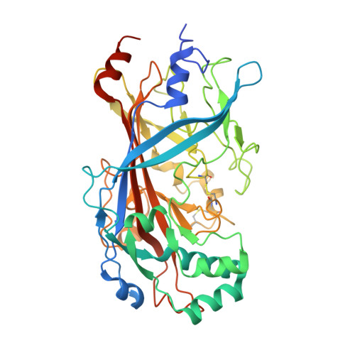
    Enterovirus 71 (EV71) is a common cause of hand, foot and mouth disease-a disease endemic especially in the Asia-Pacific region 1 . Scavenger receptor class B member 2 (SCARB2) is the major receptor of EV71, as well as several other enteroviruses responsible for hand, foot and mouth disease, and plays a key role in cell entry 2 . The isolated structures of EV71 and SCARB2 are known 3-6 , but how they interact to initiate infection is not. Here, we report the EV71-SCARB2 complex structure determined at 3.4 Å resolution using cryo-electron microscopy. This reveals that SCARB2 binds EV71 on the southern rim of the canyon, rather than across the canyon, as predicted 3,7,8 . Helices 152-163 (α5) and 183-193 (α7) of SCARB2 and the viral protein 1 (VP1) GH and VP2 EF loops of EV71 dominate the interaction, suggesting an allosteric mechanism by which receptor binding might facilitate the low-pH uncoating of the virus in the endosome/lysosome. Remarkably, many residues within the binding footprint are not conserved across SCARB2-dependent enteroviruses; however, a conserved proline and glycine seem to be key residues. Thus, although the virus maintains antigenic variability even within the receptor-binding footprint, the identification of binding 'hot spots' may facilitate the design of receptor mimic therapeutics less likely to quickly generate resistance.


  • Organizational Affiliation

    Division of Structural Biology, The Wellcome Centre for Human Genetics, University of Oxford, Oxford, UK.


Macromolecules
Find similar proteins by:  (by identity cutoff)  |  3D Structure
Entity ID: 1
MoleculeChains Sequence LengthOrganismDetailsImage
Polyprotein297Enterovirus A71Mutation(s): 0 
UniProt
Find proteins for Q66479 (Human enterovirus 71 (strain 7423/MS/87))
Explore Q66479 
Go to UniProtKB:  Q66479
Entity Groups  
Sequence Clusters30% Identity50% Identity70% Identity90% Identity95% Identity100% Identity
UniProt GroupQ66479
Sequence Annotations
Expand
  • Reference Sequence
Find similar proteins by:  (by identity cutoff)  |  3D Structure
Entity ID: 2
MoleculeChains Sequence LengthOrganismDetailsImage
Polyprotein254Enterovirus A71Mutation(s): 0 
UniProt
Find proteins for Q66479 (Human enterovirus 71 (strain 7423/MS/87))
Explore Q66479 
Go to UniProtKB:  Q66479
Entity Groups  
Sequence Clusters30% Identity50% Identity70% Identity90% Identity95% Identity100% Identity
UniProt GroupQ66479
Sequence Annotations
Expand
  • Reference Sequence
Find similar proteins by:  (by identity cutoff)  |  3D Structure
Entity ID: 3
MoleculeChains Sequence LengthOrganismDetailsImage
Polyprotein242Enterovirus A71Mutation(s): 0 
UniProt
Find proteins for Q66479 (Human enterovirus 71 (strain 7423/MS/87))
Explore Q66479 
Go to UniProtKB:  Q66479
Entity Groups  
Sequence Clusters30% Identity50% Identity70% Identity90% Identity95% Identity100% Identity
UniProt GroupQ66479
Sequence Annotations
Expand
  • Reference Sequence
Find similar proteins by:  (by identity cutoff)  |  3D Structure
Entity ID: 4
MoleculeChains Sequence LengthOrganismDetailsImage
Polyprotein58Enterovirus A71Mutation(s): 0 
UniProt
Find proteins for Q66479 (Human enterovirus 71 (strain 7423/MS/87))
Explore Q66479 
Go to UniProtKB:  Q66479
Entity Groups  
Sequence Clusters30% Identity50% Identity70% Identity90% Identity95% Identity100% Identity
UniProt GroupQ66479
Sequence Annotations
Expand
  • Reference Sequence
Find similar proteins by:  (by identity cutoff)  |  3D Structure
Entity ID: 5
MoleculeChains Sequence LengthOrganismDetailsImage
Lysosome membrane protein 2416Homo sapiensMutation(s): 0 
Gene Names: SCARB2CD36L2LIMP2LIMPII
UniProt & NIH Common Fund Data Resources
Find proteins for Q14108 (Homo sapiens)
Explore Q14108 
Go to UniProtKB:  Q14108
PHAROS:  Q14108
GTEx:  ENSG00000138760 
Entity Groups  
Sequence Clusters30% Identity50% Identity70% Identity90% Identity95% Identity100% Identity
UniProt GroupQ14108
Sequence Annotations
Expand
  • Reference Sequence
Oligosaccharides

Help

Entity ID: 6
MoleculeChains Length2D Diagram Glycosylation3D Interactions
alpha-D-mannopyranose-(1-3)-beta-D-mannopyranose-(1-4)-2-acetamido-2-deoxy-beta-D-glucopyranose-(1-4)-2-acetamido-2-deoxy-beta-D-glucopyranose
F
4N-Glycosylation
Glycosylation Resources
GlyTouCan:  G81315DD
GlyCosmos:  G81315DD
GlyGen:  G81315DD
Entity ID: 7
MoleculeChains Length2D Diagram Glycosylation3D Interactions
2-acetamido-2-deoxy-beta-D-glucopyranose-(1-4)-2-acetamido-2-deoxy-beta-D-glucopyranose
G, I
2N-Glycosylation
Glycosylation Resources
GlyTouCan:  G42666HT
GlyCosmos:  G42666HT
GlyGen:  G42666HT
Entity ID: 8
MoleculeChains Length2D Diagram Glycosylation3D Interactions
beta-D-mannopyranose-(1-4)-2-acetamido-2-deoxy-beta-D-glucopyranose-(1-4)-2-acetamido-2-deoxy-beta-D-glucopyranose
H
3N-Glycosylation
Glycosylation Resources
GlyTouCan:  G15407YE
GlyCosmos:  G15407YE
GlyGen:  G15407YE
Entity ID: 9
MoleculeChains Length2D Diagram Glycosylation3D Interactions
alpha-D-mannopyranose-(1-2)-alpha-D-mannopyranose-(1-2)-alpha-D-mannopyranose-(1-3)-[alpha-D-mannopyranose-(1-2)-alpha-D-mannopyranose-(1-3)-[alpha-D-mannopyranose-(1-2)-alpha-D-mannopyranose-(1-6)]alpha-D-mannopyranose-(1-6)]beta-D-mannopyranose-(1-4)-2-acetamido-2-deoxy-beta-D-glucopyranose-(1-4)-2-acetamido-2-deoxy-beta-D-glucopyranose
J
11N-Glycosylation
Glycosylation Resources
GlyTouCan:  G60230HH
GlyCosmos:  G60230HH
GlyGen:  G60230HH
Entity ID: 10
MoleculeChains Length2D Diagram Glycosylation3D Interactions
alpha-D-mannopyranose-(1-3)-[alpha-D-mannopyranose-(1-6)]beta-D-mannopyranose-(1-4)-2-acetamido-2-deoxy-beta-D-glucopyranose-(1-4)-2-acetamido-2-deoxy-beta-D-glucopyranose
K
5N-Glycosylation
Glycosylation Resources
GlyTouCan:  G22768VO
GlyCosmos:  G22768VO
GlyGen:  G22768VO
Small Molecules
Ligands 2 Unique
IDChains Name / Formula / InChI Key2D Diagram3D Interactions
906
Query on 906

Download Ideal Coordinates CCD File 
L [auth A]1-(2-aminopyridin-4-yl)-3-[(3S)-5-{4-[(E)-(ethoxyimino)methyl]phenoxy}-3-methylpentyl]imidazolidin-2-one
C23 H31 N5 O3
SRJKGDFZRYDLKM-WYZSWBMLSA-N
NAG
Query on NAG

Download Ideal Coordinates CCD File 
M [auth E],
N [auth E]
2-acetamido-2-deoxy-beta-D-glucopyranose
C8 H15 N O6
OVRNDRQMDRJTHS-FMDGEEDCSA-N
Experimental Data & Validation

Experimental Data

  • Method: ELECTRON MICROSCOPY
  • Resolution: 3.40 Å
  • Aggregation State: PARTICLE 
  • Reconstruction Method: SINGLE PARTICLE 
EM Software:
TaskSoftware PackageVersion
RECONSTRUCTIONRELION2
MODEL REFINEMENTPHENIX

Structure Validation

View Full Validation Report


Entry History & Funding Information

Deposition Data


Funding OrganizationLocationGrant Number
Medical Research Council (United Kingdom)United KingdomMR/N00065X/1

Revision History  (Full details and data files)

  • Version 1.0: 2018-11-28
    Type: Initial release
  • Version 1.1: 2018-12-19
    Changes: Data collection, Database references
  • Version 1.2: 2019-03-06
    Changes: Data collection, Database references
  • Version 2.0: 2020-07-29
    Type: Remediation
    Reason: Carbohydrate remediation
    Changes: Atomic model, Data collection, Derived calculations, Structure summary