6HWL

Glucosamine kinase in complex with glucosamine, ADP and inorganic phosphate


Experimental Data Snapshot

  • Method: X-RAY DIFFRACTION
  • Resolution: 2.15 Å
  • R-Value Free: 0.243 
  • R-Value Work: 0.207 
  • R-Value Observed: 0.209 

wwPDB Validation   3D Report Full Report


Ligand Structure Quality Assessment 


This is version 1.4 of the entry. See complete history


Literature

Molecular Fingerprints for a Novel Enzyme Family in Actinobacteria with Glucosamine Kinase Activity.

Manso, J.A.Nunes-Costa, D.Macedo-Ribeiro, S.Empadinhas, N.Pereira, P.J.B.

(2019) mBio 10

  • DOI: https://doi.org/10.1128/mBio.00239-19
  • Primary Citation of Related Structures:  
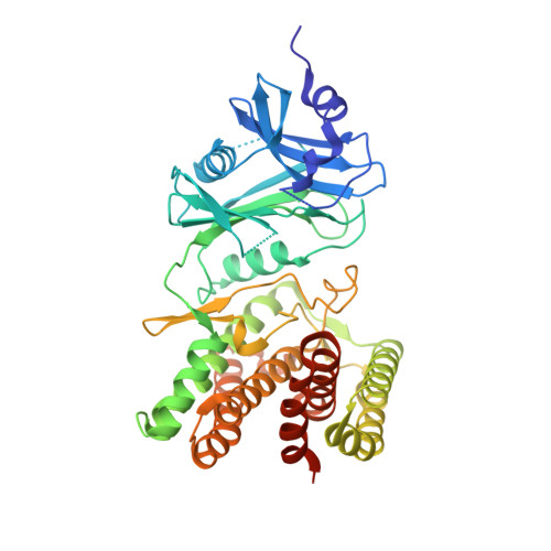
    6HWJ, 6HWK, 6HWL

  • PubMed Abstract: 

    Actinobacteria have long been the main source of antibiotics, secondary metabolites with tightly controlled biosynthesis by environmental and physiological factors. Phosphorylation of exogenous glucosamine has been suggested as a mechanism for incorporation of this extracellular material into secondary metabolite biosynthesis, but experimental evidence of specific glucosamine kinases in Actinobacteria is lacking. Here, we present the molecular fingerprints for the identification of a unique family of actinobacterial glucosamine kinases. Structural and biochemical studies on a distinctive kinase from the soil bacterium Streptacidiphilus jiangxiensis unveiled its preference for glucosamine and provided structural evidence of a phosphoryl transfer to this substrate. Conservation of glucosamine-contacting residues across a large number of uncharacterized actinobacterial proteins unveiled a specific glucosamine binding sequence motif. This family of kinases and their genetic context may represent the missing link for the incorporation of environmental glucosamine into the antibiotic biosynthesis pathways in Actinobacteria and can be explored to enhance antibiotic production. IMPORTANCE The discovery of novel enzymes involved in antibiotic biosynthesis pathways is currently a topic of utmost importance. The high levels of antibiotic resistance detected worldwide threaten our ability to combat infections and other 20th-century medical achievements, namely, organ transplantation or cancer chemotherapy. We have identified and characterized a unique family of enzymes capable of phosphorylating glucosamine to glucosamine-6-phosphate, a crucial molecule directly involved in the activation of antibiotic production pathways in Actinobacteria , nature's main source of antimicrobials. The consensus sequence identified for these glucosamine kinases will help establish a molecular fingerprint to reveal yet-uncharacterized sequences in antibiotic producers, which should have an important impact in biotechnological and biomedical applications, including the enhancement and optimization of antibiotic production.


  • Organizational Affiliation

    IBMC-Instituto de Biologia Molecular e Celular, Universidade do Porto, Porto, Portugal.


Macromolecules
Find similar proteins by:  (by identity cutoff)  |  3D Structure
Entity ID: 1
MoleculeChains Sequence LengthOrganismDetailsImage
Glucosamine kinase
A, B
451Streptacidiphilus jiangxiensisMutation(s): 0 
Gene Names: SAMN05414137_114149
UniProt
Find proteins for A0A1H7TQR5 (Streptacidiphilus jiangxiensis)
Explore A0A1H7TQR5 
Go to UniProtKB:  A0A1H7TQR5
Entity Groups  
Sequence Clusters30% Identity50% Identity70% Identity90% Identity95% Identity100% Identity
UniProt GroupA0A1H7TQR5
Sequence Annotations
Expand
  • Reference Sequence
Small Molecules
Ligands 7 Unique
IDChains Name / Formula / InChI Key2D Diagram3D Interactions
ADP
Query on ADP

Download Ideal Coordinates CCD File 
I [auth B]ADENOSINE-5'-DIPHOSPHATE
C10 H15 N5 O10 P2
XTWYTFMLZFPYCI-KQYNXXCUSA-N
BTB
Query on BTB

Download Ideal Coordinates CCD File 
D [auth A]2-[BIS-(2-HYDROXY-ETHYL)-AMINO]-2-HYDROXYMETHYL-PROPANE-1,3-DIOL
C8 H19 N O5
OWMVSZAMULFTJU-UHFFFAOYSA-N
GCS
Query on GCS

Download Ideal Coordinates CCD File 
M [auth B]2-amino-2-deoxy-beta-D-glucopyranose
C6 H13 N O5
MSWZFWKMSRAUBD-QZABAPFNSA-N
PEG
Query on PEG

Download Ideal Coordinates CCD File 
E [auth A],
F [auth A],
G [auth A]
DI(HYDROXYETHYL)ETHER
C4 H10 O3
MTHSVFCYNBDYFN-UHFFFAOYSA-N
PO4
Query on PO4

Download Ideal Coordinates CCD File 
L [auth B]PHOSPHATE ION
O4 P
NBIIXXVUZAFLBC-UHFFFAOYSA-K
CL
Query on CL

Download Ideal Coordinates CCD File 
C [auth A],
H [auth B]
CHLORIDE ION
Cl
VEXZGXHMUGYJMC-UHFFFAOYSA-M
MG
Query on MG

Download Ideal Coordinates CCD File 
J [auth B],
K [auth B]
MAGNESIUM ION
Mg
JLVVSXFLKOJNIY-UHFFFAOYSA-N
Experimental Data & Validation

Experimental Data

Unit Cell:
Length ( Å )Angle ( ˚ )
a = 58.764α = 90
b = 97.435β = 107.42
c = 80.188γ = 90
Software Package:
Software NamePurpose
PHENIXrefinement
XDSdata reduction
autoPROCdata scaling
PHASERphasing

Structure Validation

View Full Validation Report


Ligand Structure Quality Assessment 


Entry History 

Deposition Data

Revision History  (Full details and data files)

  • Version 1.0: 2019-05-01
    Type: Initial release
  • Version 1.1: 2019-05-22
    Changes: Data collection, Database references
  • Version 1.2: 2020-07-08
    Changes: Data collection
  • Version 1.3: 2020-07-29
    Type: Remediation
    Reason: Carbohydrate remediation
    Changes: Data collection, Derived calculations, Refinement description, Structure summary
  • Version 1.4: 2020-09-30
    Changes: Database references, Structure summary