6HTT

Crystal structure of Schistosoma mansoni HDAC8 complexed with a benzohydroxamate inhibitor 7


Experimental Data Snapshot

  • Method: X-RAY DIFFRACTION
  • Resolution: 1.75 Å
  • R-Value Free: 0.183 
  • R-Value Work: 0.150 
  • R-Value Observed: 0.151 

wwPDB Validation   3D Report Full Report


Ligand Structure Quality Assessment 


This is version 1.3 of the entry. See complete history


Literature

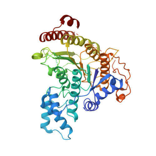
Characterization of Histone Deacetylase 8 (HDAC8) Selective Inhibition Reveals Specific Active Site Structural and Functional Determinants.

Marek, M.Shaik, T.B.Heimburg, T.Chakrabarti, A.Lancelot, J.Ramos-Morales, E.Da Veiga, C.Kalinin, D.Melesina, J.Robaa, D.Schmidtkunz, K.Suzuki, T.Holl, R.Ennifar, E.Pierce, R.J.Jung, M.Sippl, W.Romier, C.

(2018) J Med Chem 61: 10000-10016

  • DOI: https://doi.org/10.1021/acs.jmedchem.8b01087
  • Primary Citation of Related Structures:  
    6HQY, 6HRQ, 6HSF, 6HSG, 6HSH, 6HSK, 6HSZ, 6HT8, 6HTG, 6HTH, 6HTI, 6HTT, 6HTZ, 6HU0, 6HU1, 6HU2, 6HU3

  • PubMed Abstract: 

    Metal-dependent histone deacetylases (HDACs) are key epigenetic regulators that represent promising therapeutic targets for the treatment of numerous human diseases. Yet the currently FDA-approved HDAC inhibitors nonspecifically target at least several of the 11 structurally similar but functionally different HDAC isozymes, which hampers their broad usage in clinical settings. Selective inhibitors targeting single HDAC isozymes are being developed, but precise understanding in molecular terms of their selectivity remains sparse. Here, we show that HDAC8-selective inhibitors adopt a L-shaped conformation required for their binding to a HDAC8-specific pocket formed by HDAC8 catalytic tyrosine and HDAC8 L1 and L6 loops. In other HDAC isozymes, a L1-L6 lock sterically prevents L-shaped inhibitor binding. Shielding of the HDAC8-specific pocket by protein engineering decreases potency of HDAC8-selective inhibitors and affects catalytic activity. Collectively, our results unravel key HDAC8 active site structural and functional determinants important for the design of next-generation chemical probes and epigenetic drugs.


  • Organizational Affiliation

    Institut de Génétique et Biologie Moléculaire et Cellulaire (IGBMC), Département de Biologie Structurale Intégrative , Université de Strasbourg, Centre National de la Recherche Scientifique (CNRS UMR7104), Institut National de la Santé et de la Recherche Médicale (INSERM U1258) , 1 rue Laurent Fries , 67404 Illkirch Cedex , France.


Macromolecules
Find similar proteins by:  (by identity cutoff)  |  3D Structure
Entity ID: 1
MoleculeChains Sequence LengthOrganismDetailsImage
Histone deacetylase
A, B, C, D
447Schistosoma mansoniMutation(s): 0 
Gene Names: HDAC8
EC: 3.5.1.98
UniProt
Find proteins for A5H660 (Schistosoma mansoni)
Explore A5H660 
Go to UniProtKB:  A5H660
Entity Groups  
Sequence Clusters30% Identity50% Identity70% Identity90% Identity95% Identity100% Identity
UniProt GroupA5H660
Sequence Annotations
Expand
  • Reference Sequence
Small Molecules
Ligands 5 Unique
IDChains Name / Formula / InChI Key2D Diagram3D Interactions
GQZ
Query on GQZ

Download Ideal Coordinates CCD File 
CB [auth D],
H [auth A],
IA [auth C],
T [auth B]
~{N}1-oxidanyl-~{N}3-phenyl-benzene-1,3-dicarboxamide
C14 H12 N2 O3
NXHPVXNYPQJIOB-UHFFFAOYSA-N
GOL
Query on GOL

Download Ideal Coordinates CCD File 
BA [auth B]
CA [auth B]
DA [auth B]
EA [auth B]
L [auth A]
BA [auth B],
CA [auth B],
DA [auth B],
EA [auth B],
L [auth A],
LB [auth D],
M [auth A],
MB [auth D],
N [auth A],
NB [auth D],
O [auth A],
OB [auth D],
P [auth A],
RA [auth C],
SA [auth C],
TA [auth C],
UA [auth C],
VA [auth C],
WA [auth C],
XA [auth C]
GLYCEROL
C3 H8 O3
PEDCQBHIVMGVHV-UHFFFAOYSA-N
DMF
Query on DMF

Download Ideal Coordinates CCD File 
AA [auth B]
DB [auth D]
EB [auth D]
FB [auth D]
GB [auth D]
AA [auth B],
DB [auth D],
EB [auth D],
FB [auth D],
GB [auth D],
HB [auth D],
I [auth A],
IB [auth D],
J [auth A],
JA [auth C],
JB [auth D],
K [auth A],
KA [auth C],
KB [auth D],
LA [auth C],
MA [auth C],
NA [auth C],
OA [auth C],
PA [auth C],
QA [auth C],
U [auth B],
V [auth B],
W [auth B],
X [auth B],
Y [auth B],
YA [auth D],
Z [auth B]
DIMETHYLFORMAMIDE
C3 H7 N O
ZMXDDKWLCZADIW-UHFFFAOYSA-N
ZN
Query on ZN

Download Ideal Coordinates CCD File 
E [auth A],
FA [auth C],
Q [auth B],
ZA [auth D]
ZINC ION
Zn
PTFCDOFLOPIGGS-UHFFFAOYSA-N
K
Query on K

Download Ideal Coordinates CCD File 
AB [auth D]
BB [auth D]
F [auth A]
G [auth A]
GA [auth C]
AB [auth D],
BB [auth D],
F [auth A],
G [auth A],
GA [auth C],
HA [auth C],
R [auth B],
S [auth B]
POTASSIUM ION
K
NPYPAHLBTDXSSS-UHFFFAOYSA-N
Binding Affinity Annotations 
IDSourceBinding Affinity
GQZ Binding MOAD:  6HTT IC50: 220 (nM) from 1 assay(s)
Experimental Data & Validation

Experimental Data

  • Method: X-RAY DIFFRACTION
  • Resolution: 1.75 Å
  • R-Value Free: 0.183 
  • R-Value Work: 0.150 
  • R-Value Observed: 0.151 
  • Space Group: P 1
Unit Cell:
Length ( Å )Angle ( ˚ )
a = 70.4α = 75.8
b = 70.5β = 77.71
c = 98.33γ = 85.53
Software Package:
Software NamePurpose
PHENIXrefinement
XDSdata reduction
XDSdata scaling
PHENIXphasing

Structure Validation

View Full Validation Report


Ligand Structure Quality Assessment 


Entry History & Funding Information

Deposition Data


Funding OrganizationLocationGrant Number
European CommissionFrance241865
European CommissionFrance602080

Revision History  (Full details and data files)

  • Version 1.0: 2018-10-31
    Type: Initial release
  • Version 1.1: 2018-11-07
    Changes: Data collection, Database references
  • Version 1.2: 2018-11-28
    Changes: Data collection, Database references
  • Version 1.3: 2024-01-24
    Changes: Data collection, Database references, Derived calculations, Refinement description