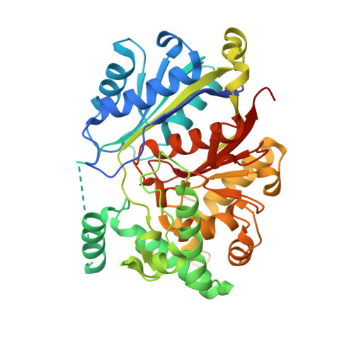
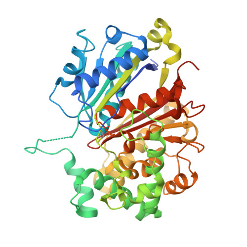
The peroxisomal zebrafish SCP2-thiolase (type-1) is a weak transient dimer as revealed by crystal structures and native mass spectrometry.
Kiema, T.R., Thapa, C.J., Laitaoja, M., Schmitz, W., Maksimainen, M.M., Fukao, T., Rouvinen, J., Janis, J., Wierenga, R.K.(2019) Biochem J 476: 307-332
- PubMed: 30573650
- DOI: https://doi.org/10.1042/BCJ20180788
- Primary Citation of Related Structures:
6HRV, 6HSJ, 6HSP - PubMed Abstract:
The SCP2 (sterol carrier protein 2)-thiolase (type-1) functions in the vertebrate peroxisomal, bile acid synthesis pathway, converting 24-keto-THC-CoA and CoA into choloyl-CoA and propionyl-CoA. This conversion concerns the β-oxidation chain shortening of the steroid fatty acyl-moiety of 24-keto-THC-CoA. This class of dimeric thiolases has previously been poorly characterized. High-resolution crystal structures of the zebrafish SCP2-thiolase (type-1) now reveal an open catalytic site, shaped by residues of both subunits. The structure of its non-dimerized monomeric form has also been captured in the obtained crystals. Four loops at the dimer interface adopt very different conformations in the monomeric form. These loops also shape the active site and their structural changes explain why a competent active site is not present in the monomeric form. Native mass spectrometry studies confirm that the zebrafish SCP2-thiolase (type-1) as well as its human homolog are weak transient dimers in solution. The crystallographic binding studies reveal the mode of binding of CoA and octanoyl-CoA in the active site, highlighting the conserved geometry of the nucleophilic cysteine, the catalytic acid/base cysteine and the two oxyanion holes. The dimer interface of SCP2-thiolase (type-1) is equally extensive as in other thiolase dimers; however, it is more polar than any of the corresponding interfaces, which correlates with the notion that the enzyme forms a weak transient dimer. The structure comparison of the monomeric and dimeric forms suggests functional relevance of this property. These comparisons provide also insights into the structural rearrangements that occur when the folded inactive monomers assemble into the mature dimer.
Organizational Affiliation:
Biocenter Oulu and Faculty of Biochemistry and Molecular Medicine, University of Oulu, PO Box 5400, FI-90014 Oulu, Finland.