


Crystal structure of the Tudor domain of human ERCC6-L2
Newman, J.A., Gavard, A.E., Nathan, W.J., Pinkas, D.M., von Delft, F., Arrowsmith, C.H., Edwards, A., Bountra, C., Gileadi, O.To be published.
Experimental Data Snapshot
wwPDB Validation 3D Report Full Report

Entity ID: 1 | |||||
|---|---|---|---|---|---|
| Molecule | Chains | Sequence Length | Organism | Details | Image |
| DNA excision repair protein ERCC-6-like 2 | 69 | Homo sapiens | Mutation(s): 0 Gene Names: ERCC6L2, C9orf102, RAD26L EC: 3.6.4 | 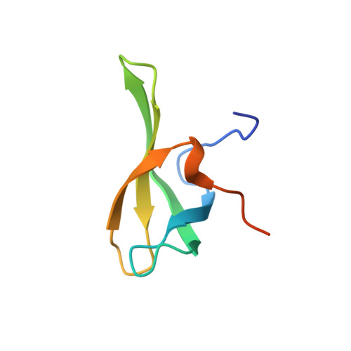 | |
UniProt & NIH Common Fund Data Resources | |||||
Find proteins for Q5T890 (Homo sapiens) Explore Q5T890 Go to UniProtKB: Q5T890 | |||||
PHAROS: Q5T890 GTEx: ENSG00000182150 | |||||
Entity Groups | |||||
| Sequence Clusters | 30% Identity50% Identity70% Identity90% Identity95% Identity100% Identity | ||||
| UniProt Group | Q5T890 | ||||
Sequence AnnotationsExpand | |||||
| |||||
| Length ( Å ) | Angle ( ˚ ) |
|---|---|
| a = 23.539 | α = 90 |
| b = 54.399 | β = 90 |
| c = 108.825 | γ = 90 |
| Software Name | Purpose |
|---|---|
| PHENIX | refinement |
| DIALS | data reduction |
| Aimless | data scaling |
| PHASER | phasing |
| Funding Organization | Location | Grant Number |
|---|---|---|
| Wellcome Trust | United Kingdom | -- |
RCSB PDB (citation) is hosted by
RCSB PDB is a member of the

