6HPF

Structure of Inactive E165Q mutant of fungal non-CBM carrying GH26 endo-b-mannanase from Yunnania penicillata in complex with alpha-62-61-di-galactosyl-mannotriose


Experimental Data Snapshot

  • Method: X-RAY DIFFRACTION
  • Resolution: 1.36 Å
  • R-Value Free: 0.143 
  • R-Value Work: 0.122 
  • R-Value Observed: 0.123 

wwPDB Validation   3D Report Full Report


Ligand Structure Quality Assessment 


This is version 2.1 of the entry. See complete history


Literature

Crystal structure and substrate interactions of an unusual fungal non-CBM carrying GH26 endo-beta-mannanase from Yunnania penicillata.

von Freiesleben, P.Moroz, O.V.Blagova, E.Wiemann, M.Spodsberg, N.Agger, J.W.Davies, G.J.Wilson, K.S.Stalbrand, H.Meyer, A.S.Krogh, K.B.R.M.

(2019) Sci Rep 9: 2266-2266

  • DOI: https://doi.org/10.1038/s41598-019-38602-x
  • Primary Citation of Related Structures:  
    6HPF

  • PubMed Abstract: 

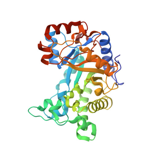
    Endo-β(1 → 4)-mannanases (endomannanases) catalyse degradation of β-mannans, an abundant class of plant polysaccharides. This study investigates structural features and substrate binding of YpenMan26A, a non-CBM carrying endomannanase from Yunnania penicillata. Structural and sequence comparisons to other fungal family GH26 endomannanases showed high sequence similarities and conserved binding residues, indicating that fungal GH26 endomannanases accommodate galactopyranosyl units in the -3 and -2 subsites. Two striking amino acid differences in the active site were found when the YpenMan26A structure was compared to a homology model of Wsp.Man26A from Westerdykella sp. and the sequences of nine other fungal GH26 endomannanases. Two YpenMan26A mutants, W110H and D37T, inspired by differences observed in Wsp.Man26A, produced a shift in how mannopentaose bound across the active site cleft and a decreased affinity for galactose in the -2 subsite, respectively, compared to YpenMan26A. YpenMan26A was moreover found to have a flexible surface loop in the position where PansMan26A from Podospora anserina has an α-helix (α9) which interacts with its family 35 CBM. Sequence alignment inferred that the core structure of fungal GH26 endomannanases differ depending on the natural presence of this type of CBM. These new findings have implications for selecting and optimising these enzymes for galactomannandegradation.


  • Organizational Affiliation

    Novozymes A/S, Krogshøjvej 36, 2880, Bagsværd, Denmark.


Macromolecules
Find similar proteins by:  (by identity cutoff)  |  3D Structure
Entity ID: 1
MoleculeChains Sequence LengthOrganismDetailsImage
endo-b-mannanase312Yunnania penicillataMutation(s): 0 
UniProt
Find proteins for A0A3G4RHU4 (Yunnania penicillata)
Explore A0A3G4RHU4 
Go to UniProtKB:  A0A3G4RHU4
Entity Groups  
Sequence Clusters30% Identity50% Identity70% Identity90% Identity95% Identity100% Identity
UniProt GroupA0A3G4RHU4
Sequence Annotations
Expand
  • Reference Sequence
Oligosaccharides

Help

Entity ID: 2
MoleculeChains Length2D Diagram Glycosylation3D Interactions
beta-D-mannopyranose-(1-4)-[alpha-D-galactopyranose-(1-6)]beta-D-mannopyranose-(1-4)-[alpha-D-galactopyranose-(1-6)]alpha-D-mannopyranose
B
5N/A
Glycosylation Resources
GlyTouCan:  G29689VG
GlyCosmos:  G29689VG
Small Molecules
Ligands 4 Unique
IDChains Name / Formula / InChI Key2D Diagram3D Interactions
NAG
Query on NAG

Download Ideal Coordinates CCD File 
D [auth A]2-acetamido-2-deoxy-beta-D-glucopyranose
C8 H15 N O6
OVRNDRQMDRJTHS-FMDGEEDCSA-N
SO4
Query on SO4

Download Ideal Coordinates CCD File 
E [auth A]
F [auth A]
G [auth A]
H [auth A]
I [auth A]
E [auth A],
F [auth A],
G [auth A],
H [auth A],
I [auth A],
J [auth A]
SULFATE ION
O4 S
QAOWNCQODCNURD-UHFFFAOYSA-L
ACY
Query on ACY

Download Ideal Coordinates CCD File 
C [auth A]ACETIC ACID
C2 H4 O2
QTBSBXVTEAMEQO-UHFFFAOYSA-N
CL
Query on CL

Download Ideal Coordinates CCD File 
K [auth A]CHLORIDE ION
Cl
VEXZGXHMUGYJMC-UHFFFAOYSA-M
Experimental Data & Validation

Experimental Data

  • Method: X-RAY DIFFRACTION
  • Resolution: 1.36 Å
  • R-Value Free: 0.143 
  • R-Value Work: 0.122 
  • R-Value Observed: 0.123 
  • Space Group: P 65 2 2
Unit Cell:
Length ( Å )Angle ( ˚ )
a = 98.988α = 90
b = 98.988β = 90
c = 170.497γ = 120
Software Package:
Software NamePurpose
REFMACrefinement
xia2data reduction
Aimlessdata scaling
MOLREPphasing

Structure Validation

View Full Validation Report


Ligand Structure Quality Assessment 


Entry History 

Revision History  (Full details and data files)

  • Version 1.0: 2019-03-06
    Type: Initial release
  • Version 2.0: 2020-07-29
    Type: Remediation
    Reason: Carbohydrate remediation
    Changes: Atomic model, Data collection, Derived calculations, Structure summary
  • Version 2.1: 2024-01-24
    Changes: Data collection, Database references, Derived calculations, Refinement description, Structure summary