6HF1

Mutant oxidoreductase fragment of mouse QSOX1 in complex with an antibody Fab


Experimental Data Snapshot

  • Method: X-RAY DIFFRACTION
  • Resolution: 1.94 Å
  • R-Value Free: 0.231 
  • R-Value Work: 0.194 
  • R-Value Observed: 0.196 

wwPDB Validation   3D Report Full Report


This is version 1.1 of the entry. See complete history


Literature

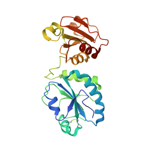
cis-Proline mutants of quiescin sulfhydryl oxidase 1 with altered redox properties undermine extracellular matrix integrity and cell adhesion in fibroblast cultures.

Javitt, G.Grossman-Haham, I.Alon, A.Resnick, E.Mutsafi, Y.Ilani, T.Fass, D.

(2019) Protein Sci 28: 228-238

  • DOI: https://doi.org/10.1002/pro.3537
  • Primary Citation of Related Structures:  
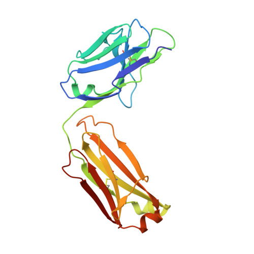
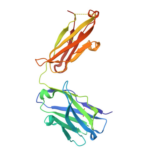
    6HF1

  • PubMed Abstract: 

    The thioredoxin superfamily has expanded and diverged extensively throughout evolution such that distant members no longer show appreciable sequence homology. Nevertheless, redox-active thioredoxin-fold proteins functioning in diverse physiological contexts often share canonical amino acids near the active-site (di-)cysteine motif. Quiescin sulfhydryl oxidase 1 (QSOX1), a catalyst of disulfide bond formation secreted by fibroblasts, is a multi-domain thioredoxin superfamily enzyme with certain similarities to the protein disulfide isomerase (PDI) enzymes. Among other potential functions, QSOX1 supports extracellular matrix assembly in fibroblast cultures. We introduced mutations at a cis-proline in QSOX1 that is conserved across the thioredoxin superfamily and was previously observed to modulate redox interactions of the bacterial enzyme DsbA. The resulting QSOX1 variants showed a striking detrimental effect when added exogenously to fibroblasts: they severely disrupted the extracellular matrix and cell adhesion, even in the presence of naturally secreted, wild-type QSOX1. The specificity of this phenomenon for particular QSOX1 mutants inspired an investigation of the effects of mutation on catalytic and redox properties. For a series of QSOX1 mutants, the detrimental effect correlated with the redox potential of the first redox-active site, and an X-ray crystal structure of one of the mutants revealed the reorganization of the cis-proline loop caused by the mutations. Due to the conservation of the mutated residues across the PDI family and beyond, insights obtained in this study may be broadly applicable to a variety of physiologically important redox-active enzymes. IMPACT STATEMENT: We show that mutation of a conserved cis-proline amino acid, analogous to a mutation used to trap substrates of a bacterial disulfide catalyst, has a dramatic effect on the physiological function of the mammalian disulfide catalyst QSOX1. As the active-site region of QSOX1 is shared with the large family of protein disulfide isomerases in humans, the effects of such mutations on redox properties, enzymatic activity, and biological targeting may be relevant across the family.


  • Organizational Affiliation

    Department of Structural Biology, Weizmann Institute of Science, Rehovot, 7610001, Israel.


Macromolecules
Find similar proteins by:  (by identity cutoff)  |  3D Structure
Entity ID: 1
MoleculeChains Sequence LengthOrganismDetailsImage
Sulfhydryl oxidase 1
A, D
238Mus musculusMutation(s): 2 
Gene Names: Qsox1Qscn6Sox
EC: 1.8.3.2
UniProt & NIH Common Fund Data Resources
Find proteins for Q8BND5 (Mus musculus)
Explore Q8BND5 
Go to UniProtKB:  Q8BND5
IMPC:  MGI:1330818
Entity Groups  
Sequence Clusters30% Identity50% Identity70% Identity90% Identity95% Identity100% Identity
UniProt GroupQ8BND5
Sequence Annotations
Expand
  • Reference Sequence
Find similar proteins by:  (by identity cutoff)  |  3D Structure
Entity ID: 2
MoleculeChains Sequence LengthOrganismDetailsImage
Fab 316 heavy chainB [auth C]215Mus musculusMutation(s): 0 
Entity Groups  
Sequence Clusters30% Identity50% Identity70% Identity90% Identity95% Identity100% Identity
Sequence Annotations
Expand
  • Reference Sequence
Find similar proteins by:  (by identity cutoff)  |  3D Structure
Entity ID: 3
MoleculeChains Sequence LengthOrganismDetailsImage
Fab 316 light chainC [auth B],
F [auth E]
212Mus musculusMutation(s): 0 
Entity Groups  
Sequence Clusters30% Identity50% Identity70% Identity90% Identity95% Identity100% Identity
Sequence Annotations
Expand
  • Reference Sequence
Find similar proteins by:  (by identity cutoff)  |  3D Structure
Entity ID: 4
MoleculeChains Sequence LengthOrganismDetailsImage
Fab 316 heavy chainE [auth F]216Mus musculusMutation(s): 0 
Entity Groups  
Sequence Clusters30% Identity50% Identity70% Identity90% Identity95% Identity100% Identity
Sequence Annotations
Expand
  • Reference Sequence
Experimental Data & Validation

Experimental Data

  • Method: X-RAY DIFFRACTION
  • Resolution: 1.94 Å
  • R-Value Free: 0.231 
  • R-Value Work: 0.194 
  • R-Value Observed: 0.196 
  • Space Group: P 21 21 21
Unit Cell:
Length ( Å )Angle ( ˚ )
a = 65.539α = 90
b = 112.715β = 90
c = 193.418γ = 90
Software Package:
Software NamePurpose
PHENIXrefinement
PDB_EXTRACTdata extraction
HKL-3000data reduction
HKL-3000data scaling
PHENIXphasing

Structure Validation

View Full Validation Report


Entry History & Funding Information

Deposition Data


Funding OrganizationLocationGrant Number
European Research Council310649

Revision History  (Full details and data files)

  • Version 1.0: 2019-04-03
    Type: Initial release
  • Version 1.1: 2024-01-17
    Changes: Data collection, Database references, Refinement description