6HB3

Structure of Hgh1, crystal form II


Experimental Data Snapshot

  • Method: X-RAY DIFFRACTION
  • Resolution: 3.00 Å
  • R-Value Free: 0.230 
  • R-Value Work: 0.220 
  • R-Value Observed: 0.220 

wwPDB Validation   3D Report Full Report


This is version 1.2 of the entry. See complete history


Literature

Chaperone Function of Hgh1 in the Biogenesis of Eukaryotic Elongation Factor 2.

Monkemeyer, L.Klaips, C.L.Balchin, D.Korner, R.Hartl, F.U.Bracher, A.

(2019) Mol Cell 74: 88-100.e9

  • DOI: https://doi.org/10.1016/j.molcel.2019.01.034
  • Primary Citation of Related Structures:  
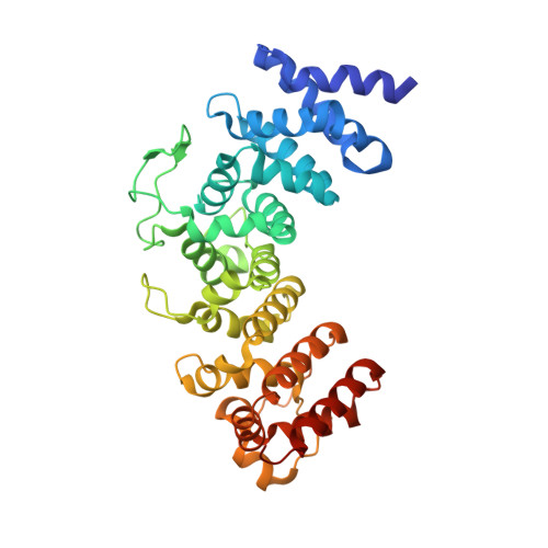
    6HB1, 6HB2, 6HB3

  • PubMed Abstract: 

    Eukaryotic elongation factor 2 (eEF2) is an abundant and essential component of the translation machinery. The biogenesis of this 93 kDa multi-domain protein is assisted by the chaperonin TRiC/CCT. Here, we show in yeast cells that the highly conserved protein Hgh1 (FAM203 in humans) is a chaperone that cooperates with TRiC in eEF2 folding. In the absence of Hgh1, a substantial fraction of newly synthesized eEF2 is degraded or aggregates. We solved the crystal structure of Hgh1 and analyzed the interaction of wild-type and mutant Hgh1 with eEF2. These experiments revealed that Hgh1 is an armadillo repeat protein that binds to the dynamic central domain III of eEF2 via a bipartite interface. Hgh1 binding recruits TRiC to the C-terminal eEF2 module and prevents unproductive interactions of domain III, allowing efficient folding of the N-terminal GTPase module. eEF2 folding is completed upon dissociation of TRiC and Hgh1.


  • Organizational Affiliation

    Department of Cellular Biochemistry, Max Planck Institute of Biochemistry, Am Klopferspitz 18, 82152 Martinsried, Germany.


Macromolecules
Find similar proteins by:  (by identity cutoff)  |  3D Structure
Entity ID: 1
MoleculeChains Sequence LengthOrganismDetailsImage
Protein HGH1
A, B, C, D
369Saccharomyces cerevisiae S288CMutation(s): 0 
Gene Names: HGH1YGR187CG7538
UniProt
Find proteins for P48362 (Saccharomyces cerevisiae (strain ATCC 204508 / S288c))
Explore P48362 
Go to UniProtKB:  P48362
Entity Groups  
Sequence Clusters30% Identity50% Identity70% Identity90% Identity95% Identity100% Identity
UniProt GroupP48362
Sequence Annotations
Expand
  • Reference Sequence
Experimental Data & Validation

Experimental Data

  • Method: X-RAY DIFFRACTION
  • Resolution: 3.00 Å
  • R-Value Free: 0.230 
  • R-Value Work: 0.220 
  • R-Value Observed: 0.220 
  • Space Group: C 1 2 1
Unit Cell:
Length ( Å )Angle ( ˚ )
a = 191.798α = 90
b = 69.643β = 93.75
c = 206.866γ = 90
Software Package:
Software NamePurpose
XDSdata reduction
Aimlessdata scaling
MOLREPphasing
REFMACrefinement
PDB_EXTRACTdata extraction

Structure Validation

View Full Validation Report


Entry History 

Deposition Data

Revision History  (Full details and data files)

  • Version 1.0: 2019-02-27
    Type: Initial release
  • Version 1.1: 2019-03-27
    Changes: Data collection, Database references
  • Version 1.2: 2019-04-17
    Changes: Data collection, Database references