Unraveling the role of the secretor antigen in human rotavirus attachment to histo-blood group antigens.
Gozalbo-Rovira, R., Ciges-Tomas, J.R., Vila-Vicent, S., Buesa, J., Santiso-Bellon, C., Monedero, V., Yebra, M.J., Marina, A., Rodriguez-Diaz, J.(2019) PLoS Pathog 15: e1007865-e1007865
- PubMed: 31226167
- DOI: https://doi.org/10.1371/journal.ppat.1007865
- Primary Citation of Related Structures:
6H9W, 6H9Z - PubMed Abstract:
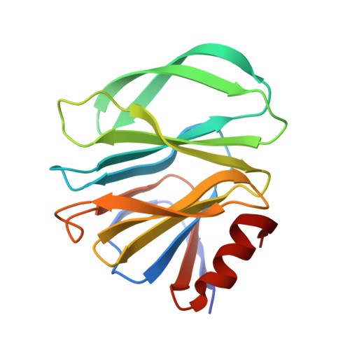
Rotavirus is the leading agent causing acute gastroenteritis in young children, with the P[8] genotype accounting for more than 80% of infections in humans. The molecular bases for binding of the VP8* domain from P[8] VP4 spike protein to its cellular receptor, the secretory H type-1 antigen (Fuc-α1,2-Gal-β1,3-GlcNAc; H1), and to its precursor lacto-N-biose (Gal-β1,3-GlcNAc; LNB) have been determined. The resolution of P[8] VP8* crystal structures in complex with H1 antigen and LNB and site-directed mutagenesis experiments revealed that both glycans bind to the P[8] VP8* protein through a binding pocket shared with other members of the P[II] genogroup (i.e.: P[4], P[6] and P[19]). Our results show that the L-fucose moiety from H1 only displays indirect contacts with P[8] VP8*. However, the induced conformational changes in the LNB moiety increase the ligand affinity by two-fold, as measured by surface plasmon resonance (SPR), providing a molecular explanation for the different susceptibility to rotavirus infection between secretor and non-secretor individuals. The unexpected interaction of P[8] VP8* with LNB, a building block of type-1 human milk oligosaccharides, resulted in inhibition of rotavirus infection, highlighting the role and possible application of this disaccharide as an antiviral. While key amino acids in the H1/LNB binding pocket were highly conserved in members of the P[II] genogroup, differences were found in ligand affinities among distinct P[8] genetic lineages. The variation in affinities were explained by subtle structural differences induced by amino acid changes in the vicinity of the binding pocket, providing a fine-tuning mechanism for glycan binding in P[8] rotavirus.
Organizational Affiliation:
Departament of Microbiology, Faculty of Medicine, University of Valencia, Valencia, Spain.