6GXV

Amylase in complex with acarbose


Experimental Data Snapshot

  • Method: X-RAY DIFFRACTION
  • Resolution: 2.07 Å
  • R-Value Free: 0.176 
  • R-Value Work: 0.138 
  • R-Value Observed: 0.140 

wwPDB Validation   3D Report Full Report


Ligand Structure Quality Assessment 


This is version 2.1 of the entry. See complete history


Literature

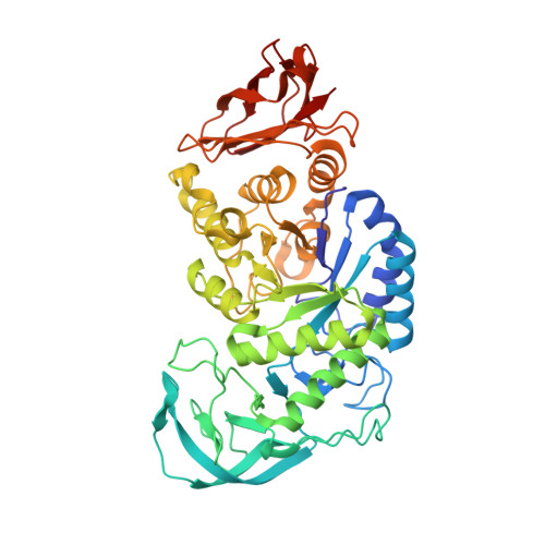
The structure of the AliC GH13 alpha-amylase from Alicyclobacillus sp. reveals the accommodation of starch branching points in the alpha-amylase family.

Agirre, J.Moroz, O.Meier, S.Brask, J.Munch, A.Hoff, T.Andersen, C.Wilson, K.S.Davies, G.J.

(2019) Acta Crystallogr D Struct Biol 75: 1-7

  • DOI: https://doi.org/10.1107/S2059798318014900
  • Primary Citation of Related Structures:  
    6GXV, 6GYA

  • PubMed Abstract: 

    α-Amylases are glycoside hydrolases that break the α-1,4 bonds in starch and related glycans. The degradation of starch is rendered difficult by the presence of varying degrees of α-1,6 branch points and their possible accommodation within the active centre of α-amylase enzymes. Given the myriad industrial uses for starch and thus also for α-amylase-catalysed starch degradation and modification, there is considerable interest in how different α-amylases might accommodate these branches, thus impacting on the potential processing of highly branched post-hydrolysis remnants (known as limit dextrins) and societal applications. Here, it was sought to probe the branch-point accommodation of the Alicyclobacillus sp. CAZy family GH13 α-amylase AliC, prompted by the observation of a molecule of glucose in a position that may represent a branch point in an acarbose complex solved at 2.1 Å resolution. Limit digest analysis by two-dimensional NMR using both pullulan (a regular linear polysaccharide of α-1,4, α-1,4, α-1,6 repeating trisaccharides) and amylopectin starch showed how the Alicyclobacillus sp. enzyme could accept α-1,6 branches in at least the -2, +1 and +2 subsites, consistent with the three-dimensional structures with glucosyl moieties in the +1 and +2 subsites and the solvent-exposure of the -2 subsite 6-hydroxyl group. Together, the work provides a rare insight into branch-point acceptance in these industrial catalysts.


  • Organizational Affiliation

    York Structural Biology Laboratory, Department of Chemistry, The University of York, York YO10 5DD, England.


Macromolecules
Find similar proteins by:  (by identity cutoff)  |  3D Structure
Entity ID: 1
MoleculeChains Sequence LengthOrganismDetailsImage
A-amylase
A, B
484Alicyclobacillus sp.Mutation(s): 0 
UniProt
Find proteins for A0A3P8MUS3 (Alicyclobacillus sp)
Explore A0A3P8MUS3 
Go to UniProtKB:  A0A3P8MUS3
Entity Groups  
Sequence Clusters30% Identity50% Identity70% Identity90% Identity95% Identity100% Identity
UniProt GroupA0A3P8MUS3
Sequence Annotations
Expand
  • Reference Sequence
Oligosaccharides

Help

Entity ID: 2
MoleculeChains Length2D Diagram Glycosylation3D Interactions
alpha-D-glucopyranose-(1-4)-alpha-D-glucopyranose
C, D
2N/A
Glycosylation Resources
GlyTouCan:  G07411ON
GlyCosmos:  G07411ON
Entity ID: 3
MoleculeChains Length2D Diagram Glycosylation3D Interactions
4,6-dideoxy-4-{[(1S,4R,5S,6S)-4,5,6-trihydroxy-3-(hydroxymethyl)cyclohex-2-en-1-yl]amino}-alpha-D-glucopyranose-(1-4)-alpha-D-glucopyranose-(1-4)-4,6-dideoxy-4-{[(1S,4R,5S,6S)-4,5,6-trihydroxy-3-(hydroxymethyl)cyclohex-2-en-1-yl]amino}-alpha-D-glucopyranose-(1-4)-alpha-D-glucopyranose
E, F
4N/AN/A
Glycosylation Resources
GlyTouCan:  G53811MA
GlyCosmos:  G53811MA
Small Molecules
Ligands 4 Unique
IDChains Name / Formula / InChI Key2D Diagram3D Interactions
BGC
Query on BGC

Download Ideal Coordinates CCD File 
L [auth A],
N [auth A],
O [auth B],
Q [auth B]
beta-D-glucopyranose
C6 H12 O6
WQZGKKKJIJFFOK-VFUOTHLCSA-N
GLC
Query on GLC

Download Ideal Coordinates CCD File 
K [auth A],
M [auth A],
P [auth B],
V [auth B]
alpha-D-glucopyranose
C6 H12 O6
WQZGKKKJIJFFOK-DVKNGEFBSA-N
CA
Query on CA

Download Ideal Coordinates CCD File 
G [auth A]
I [auth A]
J [auth A]
R [auth B]
T [auth B]
G [auth A],
I [auth A],
J [auth A],
R [auth B],
T [auth B],
U [auth B]
CALCIUM ION
Ca
BHPQYMZQTOCNFJ-UHFFFAOYSA-N
NA
Query on NA

Download Ideal Coordinates CCD File 
H [auth A],
S [auth B]
SODIUM ION
Na
FKNQFGJONOIPTF-UHFFFAOYSA-N
Biologically Interesting Molecules (External Reference) 1 Unique
Experimental Data & Validation

Experimental Data

  • Method: X-RAY DIFFRACTION
  • Resolution: 2.07 Å
  • R-Value Free: 0.176 
  • R-Value Work: 0.138 
  • R-Value Observed: 0.140 
  • Space Group: P 41 21 2
Unit Cell:
Length ( Å )Angle ( ˚ )
a = 180.9α = 90
b = 180.9β = 90
c = 77.85γ = 90
Software Package:
Software NamePurpose
REFMACrefinement
Aimlessdata scaling
BUCCANEERmodel building
MOLREPphasing
Cootmodel building

Structure Validation

View Full Validation Report


Ligand Structure Quality Assessment 


Entry History 

Deposition Data

Revision History  (Full details and data files)

  • Version 1.0: 2019-01-23
    Type: Initial release
  • Version 1.1: 2019-05-22
    Changes: Data collection, Database references, Source and taxonomy, Structure summary
  • Version 1.2: 2019-05-29
    Changes: Data collection, Source and taxonomy
  • Version 1.3: 2019-08-21
    Changes: Data collection
  • Version 2.0: 2020-07-29
    Type: Remediation
    Reason: Carbohydrate remediation
    Changes: Atomic model, Data collection, Derived calculations, Non-polymer description, Structure summary
  • Version 2.1: 2024-01-17
    Changes: Data collection, Database references, Derived calculations, Refinement description, Structure summary