6GNI

Cryo-tomography and subtomogram averaging of Sar1-Sec23-Sec24 - fitted model.


Experimental Data Snapshot

  • Method: ELECTRON MICROSCOPY
  • Resolution: 4.90 Å
  • Aggregation State: 3D ARRAY 
  • Reconstruction Method: SUBTOMOGRAM AVERAGING 

wwPDB Validation   3D Report Full Report


This is version 1.1 of the entry. See complete history


Literature

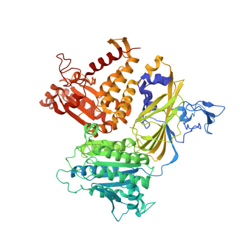
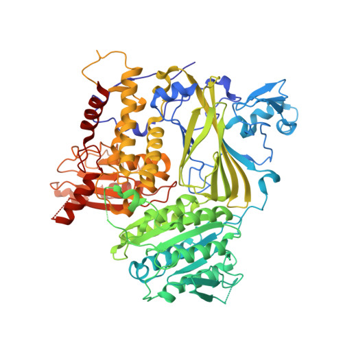
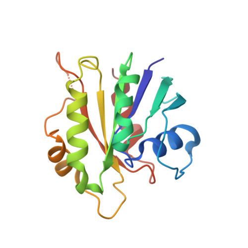
Subtomogram averaging of COPII assemblies reveals how coat organization dictates membrane shape.

Hutchings, J.Stancheva, V.Miller, E.A.Zanetti, G.

(2018) Nat Commun 9: 4154-4154

  • DOI: https://doi.org/10.1038/s41467-018-06577-4
  • Primary Citation of Related Structures:  
    6GNI

  • PubMed Abstract: 

    Eukaryotic cells employ membrane-bound carriers to transport cargo between compartments in a process essential to cell functionality. Carriers are generated by coat complexes that couple cargo capture to membrane deformation. The COPII coat mediates export from the endoplasmic reticulum by assembling in inner and outer layers, yielding carriers of variable shape and size that allow secretion of thousands of diverse cargo. Despite detailed understanding of COPII subunits, the molecular mechanisms of coat assembly and membrane deformation are unclear. Here we present a 4.9 Å cryo-tomography subtomogram averaging structure of in vitro-reconstituted membrane-bound inner coat. We show that the outer coat (Sec13-Sec31) bridges inner coat subunits (Sar1-Sec23-Sec24), promoting their assembly into a tight lattice. We directly visualize the membrane-embedded Sar1 amphipathic helix, revealing that lattice formation induces parallel helix insertions, yielding tubular curvature. We propose that regulators like the procollagen receptor TANGO1 modulate this mechanism to determine vesicle shape and size.


  • Organizational Affiliation

    Institute of Structural and Molecular Biology, Birkbeck College, Malet St., London, WC1E 7HX, UK.


Macromolecules
Find similar proteins by:  (by identity cutoff)  |  3D Structure
Entity ID: 1
MoleculeChains Sequence LengthOrganismDetailsImage
Protein transport protein SEC23767Saccharomyces cerevisiae S288CMutation(s): 0 
Gene Names: SEC23YPR181CP9705.14
UniProt
Find proteins for P15303 (Saccharomyces cerevisiae (strain ATCC 204508 / S288c))
Explore P15303 
Go to UniProtKB:  P15303
Entity Groups  
Sequence Clusters30% Identity50% Identity70% Identity90% Identity95% Identity100% Identity
UniProt GroupP15303
Sequence Annotations
Expand
  • Reference Sequence
Find similar proteins by:  (by identity cutoff)  |  3D Structure
Entity ID: 2
MoleculeChains Sequence LengthOrganismDetailsImage
Protein transport protein SEC24B [auth E]794Saccharomyces cerevisiae S288CMutation(s): 0 
Gene Names: SEC24ANU1YIL109C
UniProt
Find proteins for P40482 (Saccharomyces cerevisiae (strain ATCC 204508 / S288c))
Explore P40482 
Go to UniProtKB:  P40482
Entity Groups  
Sequence Clusters30% Identity50% Identity70% Identity90% Identity95% Identity100% Identity
UniProt GroupP40482
Sequence Annotations
Expand
  • Reference Sequence
Find similar proteins by:  (by identity cutoff)  |  3D Structure
Entity ID: 3
MoleculeChains Sequence LengthOrganismDetailsImage
Small COPII coat GTPase SAR1C [auth B]167Saccharomyces cerevisiae S288CMutation(s): 0 
Gene Names: SAR1YPL218W
EC: 3.6.5
UniProt
Find proteins for P20606 (Saccharomyces cerevisiae (strain ATCC 204508 / S288c))
Explore P20606 
Go to UniProtKB:  P20606
Entity Groups  
Sequence Clusters30% Identity50% Identity70% Identity90% Identity95% Identity100% Identity
UniProt GroupP20606
Sequence Annotations
Expand
  • Reference Sequence
Experimental Data & Validation

Experimental Data

  • Method: ELECTRON MICROSCOPY
  • Resolution: 4.90 Å
  • Aggregation State: 3D ARRAY 
  • Reconstruction Method: SUBTOMOGRAM AVERAGING 
EM Software:
TaskSoftware PackageVersion
RECONSTRUCTIONDynamo

Structure Validation

View Full Validation Report


Entry History & Funding Information

Deposition Data


Funding OrganizationLocationGrant Number
Royal SocietyUnited KingdomDH130048

Revision History  (Full details and data files)

  • Version 1.0: 2018-10-17
    Type: Initial release
  • Version 1.1: 2018-10-24
    Changes: Data collection, Database references