Structural insights into oxidation of medium-chain fatty acids and flavanone by myxobacterial cytochrome P450 CYP267B1.
Jozwik, I.K., Litzenburger, M., Khatri, Y., Schifrin, A., Girhard, M., Urlacher, V., Thunnissen, A.W.H., Bernhardt, R.(2018) Biochem J 475: 2801-2817
- PubMed: 30045877
- DOI: https://doi.org/10.1042/BCJ20180402
- Primary Citation of Related Structures:
6GK5, 6GK6 - PubMed Abstract:
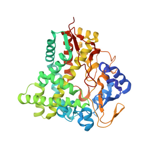
Oxidative biocatalytic reactions performed by cytochrome P450 enzymes (P450s) are of high interest for the chemical and pharmaceutical industries. CYP267B1 is a P450 enzyme from myxobacterium Sorangium cellulosum So ce56 displaying a broad substrate scope. In this work, a search for new substrates was performed, combined with product characterization and a structural analysis of substrate-bound complexes using X-ray crystallography and computational docking. The results demonstrate the ability of CYP267B1 to perform in-chain hydroxylations of medium-chain saturated fatty acids (decanoic acid, dodecanoic acid and tetradecanoic acid) and a regioselective hydroxylation of flavanone. The fatty acids are mono-hydroxylated at different in-chain positions, with decanoic acid displaying the highest regioselectivity towards ω-3 hydroxylation. Flavanone is preferably oxidized to 3-hydroxyflavanone. High-resolution crystal structures of CYP267B1 revealed a very spacious active site pocket, similarly to other P450s capable of converting macrocyclic compounds. The pocket becomes more constricted near to the heme and is closed off from solvent by residues of the F and G helices and the B-C loop. The crystal structure of the tetradecanoic acid-bound complex displays the fatty acid bound near to the heme, but in a nonproductive conformation. Molecular docking allowed modeling of the productive binding modes for the four investigated fatty acids and flavanone, as well as of two substrates identified in a previous study (diclofenac and ibuprofen), explaining the observed product profiles. The obtained structures of CYP267B1 thus serve as a valuable prediction tool for substrate hydroxylations by this highly versatile enzyme and will encourage future selectivity changes by rational protein engineering.
Organizational Affiliation:
Laboratory of Biophysical Chemistry, Groningen Biomolecular Sciences and Biotechnology Institute, University of Groningen, Nijenborgh 7, 9747 AG Groningen, The Netherlands.