6GHO

Crystal structure of Spx in complex with YjbH


Experimental Data Snapshot

  • Method: X-RAY DIFFRACTION
  • Resolution: 1.79 Å
  • R-Value Free: 0.221 
  • R-Value Work: 0.187 
  • R-Value Observed: 0.188 

wwPDB Validation   3D Report Full Report


This is version 1.1 of the entry. See complete history


Literature

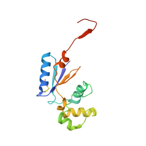
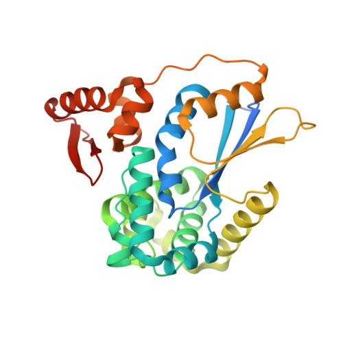
Structural Basis for YjbH Adaptor-Mediated Recognition of Transcription Factor Spx.

Awad, W.Al-Eryani, Y.Ekstrom, S.Logan, D.T.von Wachenfeldt, C.

(2019) Structure 27: 923

  • DOI: https://doi.org/10.1016/j.str.2019.03.009
  • Primary Citation of Related Structures:  
    6GHB, 6GHO

  • PubMed Abstract: 

    YjbH is a bacterial adaptor protein required for efficient proteolysis of the RNA polymerase-binding transcription factor Spx by the ClpXP protease. We report the structure of YjbH in complex with Spx. YjbH comprises a DsbA-like thioredoxin domain connected via a linker to a C-terminal domain reminiscent of the winged helix-turn-helix fold. The interaction between YjbH and Spx involves a large surface area. Binding to YjbH stabilizes the C-terminal ClpX recognition region of Spx. We show that mutation of critical YjbH contact residues abrogates Spx recognition. Small-angle X-ray scattering and hydrogen-deuterium exchange mass spectrometry analyses determined the existence of a stable heterodimeric complex in solution and provide evidence that binding of Spx to YjbH reduces the overall conformational flexibility of Spx. Our findings provide insights into the molecular basis for Spx recognition and suggest a model for how YjbH stabilizes Spx and displays the C terminus of Spx for engagement by ClpXP.


  • Organizational Affiliation

    The Microbiology Group, Department of Biology, Lund University, Sölvegatan 35, 223 62 Lund, Sweden; Department of Biophysics, Faculty of Science, Cairo University, 12316 Cairo, Egypt.


Macromolecules
Find similar proteins by:  (by identity cutoff)  |  3D Structure
Entity ID: 1
MoleculeChains Sequence LengthOrganismDetailsImage
Regulatory protein Spx132Bacillus subtilis subsp. subtilis str. 168Mutation(s): 0 
Gene Names: spxAyjbDBSU11500
UniProt
Find proteins for O31602 (Bacillus subtilis (strain 168))
Explore O31602 
Go to UniProtKB:  O31602
Entity Groups  
Sequence Clusters30% Identity50% Identity70% Identity90% Identity95% Identity100% Identity
UniProt GroupO31602
Sequence Annotations
Expand
  • Reference Sequence
Find similar proteins by:  (by identity cutoff)  |  3D Structure
Entity ID: 2
MoleculeChains Sequence LengthOrganismDetailsImage
UPF0413 protein GK0824298Geobacillus kaustophilus HTA426Mutation(s): 0 
Gene Names: GK0824
UniProt
Find proteins for Q5L1S1 (Geobacillus kaustophilus (strain HTA426))
Explore Q5L1S1 
Go to UniProtKB:  Q5L1S1
Entity Groups  
Sequence Clusters30% Identity50% Identity70% Identity90% Identity95% Identity100% Identity
UniProt GroupQ5L1S1
Sequence Annotations
Expand
  • Reference Sequence
Small Molecules
Ligands 1 Unique
IDChains Name / Formula / InChI Key2D Diagram3D Interactions
CL
Query on CL

Download Ideal Coordinates CCD File 
C [auth B]CHLORIDE ION
Cl
VEXZGXHMUGYJMC-UHFFFAOYSA-M
Experimental Data & Validation

Experimental Data

  • Method: X-RAY DIFFRACTION
  • Resolution: 1.79 Å
  • R-Value Free: 0.221 
  • R-Value Work: 0.187 
  • R-Value Observed: 0.188 
  • Space Group: P 21 21 21
Unit Cell:
Length ( Å )Angle ( ˚ )
a = 52.1α = 90
b = 85.34β = 90
c = 109.53γ = 90
Software Package:
Software NamePurpose
PHENIXrefinement
Cootmodel building
XDSdata scaling
PHENIXphasing
XDSdata reduction

Structure Validation

View Full Validation Report


Entry History 

Deposition Data

Revision History  (Full details and data files)

  • Version 1.0: 2019-04-24
    Type: Initial release
  • Version 1.1: 2019-06-19
    Changes: Data collection, Database references