6G8A

Lysozyme solved by Native SAD from a dataset collected in 5 seconds at 1 A wavelength with JUNGFRAU detector


Experimental Data Snapshot

  • Method: X-RAY DIFFRACTION
  • Resolution: 1.14 Å
  • R-Value Free: 0.130 
  • R-Value Work: 0.112 
  • R-Value Observed: 0.113 

wwPDB Validation   3D Report Full Report


This is version 1.1 of the entry. See complete history


Literature

Fast and accurate data collection for macromolecular crystallography using the JUNGFRAU detector.

Leonarski, F.Redford, S.Mozzanica, A.Lopez-Cuenca, C.Panepucci, E.Nass, K.Ozerov, D.Vera, L.Olieric, V.Buntschu, D.Schneider, R.Tinti, G.Froejdh, E.Diederichs, K.Bunk, O.Schmitt, B.Wang, M.

(2018) Nat Methods 15: 799-804

  • DOI: https://doi.org/10.1038/s41592-018-0143-7
  • Primary Citation of Related Structures:  
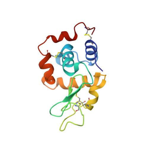
    6G89, 6G8A, 6G8B

  • PubMed Abstract: 

    The accuracy of X-ray diffraction data is directly related to how the X-ray detector records photons. Here we describe the application of a direct-detection charge-integrating pixel-array detector (JUNGFRAU) in macromolecular crystallography (MX). JUNGFRAU features a uniform response on the subpixel level, linear behavior toward high photon rates, and low-noise performance across the whole dynamic range. We demonstrate that these features allow accurate MX data to be recorded at unprecedented speed. We also demonstrate improvements over previous-generation detectors in terms of data quality, using native single-wavelength anomalous diffraction (SAD) phasing, for thaumatin, lysozyme, and aminopeptidase N. Our results suggest that the JUNGFRAU detector will substantially improve the performance of synchrotron MX beamlines and equip them for future synchrotron light sources.


  • Organizational Affiliation

    Swiss Light Source, Paul Scherrer Institute, Villigen, Switzerland.


Macromolecules
Find similar proteins by:  (by identity cutoff)  |  3D Structure
Entity ID: 1
MoleculeChains Sequence LengthOrganismDetailsImage
Lysozyme CA [auth C]129Gallus gallusMutation(s): 0 
EC: 3.2.1.17
UniProt
Find proteins for P00698 (Gallus gallus)
Explore P00698 
Go to UniProtKB:  P00698
Entity Groups  
Sequence Clusters30% Identity50% Identity70% Identity90% Identity95% Identity100% Identity
UniProt GroupP00698
Sequence Annotations
Expand
  • Reference Sequence
Small Molecules
Ligands 3 Unique
IDChains Name / Formula / InChI Key2D Diagram3D Interactions
EDO
Query on EDO

Download Ideal Coordinates CCD File 
T [auth C],
U [auth C],
V [auth C]
1,2-ETHANEDIOL
C2 H6 O2
LYCAIKOWRPUZTN-UHFFFAOYSA-N
CL
Query on CL

Download Ideal Coordinates CCD File 
B [auth C]
C
D [auth C]
E [auth C]
F [auth C]
B [auth C],
C,
D [auth C],
E [auth C],
F [auth C],
G [auth C],
H [auth C],
I [auth C],
J [auth C],
K [auth C],
L [auth C],
M [auth C],
N [auth C],
O [auth C],
P [auth C],
Q [auth C],
R [auth C]
CHLORIDE ION
Cl
VEXZGXHMUGYJMC-UHFFFAOYSA-M
NA
Query on NA

Download Ideal Coordinates CCD File 
S [auth C]SODIUM ION
Na
FKNQFGJONOIPTF-UHFFFAOYSA-N
Experimental Data & Validation

Experimental Data

  • Method: X-RAY DIFFRACTION
  • Resolution: 1.14 Å
  • R-Value Free: 0.130 
  • R-Value Work: 0.112 
  • R-Value Observed: 0.113 
  • Space Group: P 43 21 2
Unit Cell:
Length ( Å )Angle ( ˚ )
a = 78.903α = 90
b = 78.903β = 90
c = 36.941γ = 90
Software Package:
Software NamePurpose
PHENIXrefinement
XDSdata reduction
XDSdata scaling
SHELXCDphasing
SHELXEmodel building
CRANK2phasing

Structure Validation

View Full Validation Report


Entry History 

Deposition Data

Revision History  (Full details and data files)

  • Version 1.0: 2018-08-01
    Type: Initial release
  • Version 1.1: 2018-10-24
    Changes: Data collection, Database references