6G6I

MamM CTD W247A


Experimental Data Snapshot

  • Method: X-RAY DIFFRACTION
  • Resolution: 2.40 Å
  • R-Value Free: 0.262 
  • R-Value Work: 0.221 
  • R-Value Observed: 0.226 

wwPDB Validation   3D Report Full Report


This is version 1.3 of the entry. See complete history


Literature

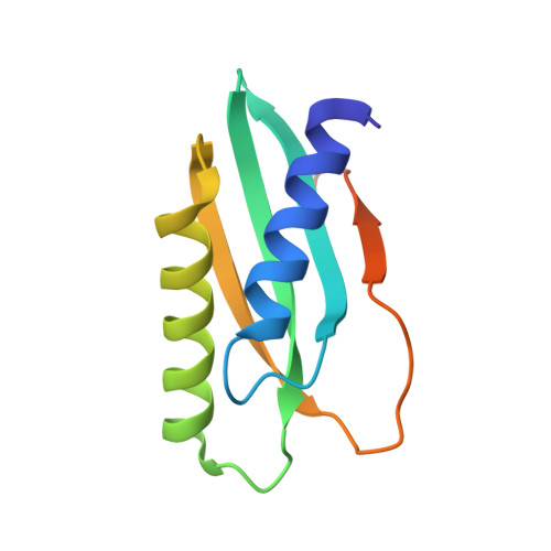
Metal binding to the dynamic cytoplasmic domain of the cation diffusion facilitator (CDF) protein MamM induces a 'locked-in' configuration.

Barber-Zucker, S.Hall, J.Mangapuram, S.V.Kass, I.Kolusheva, S.MacMillan, F.Zarivach, R.Henn, A.

(2019) FEBS J 286: 2193-2215

  • DOI: https://doi.org/10.1111/febs.14795
  • Primary Citation of Related Structures:  
    6G55, 6G5E, 6G64, 6G6I

  • PubMed Abstract: 

    Cation diffusion facilitator (CDF) proteins are a conserved family of transmembrane transporters that ensure cellular homeostasis of divalent transition metal cations. Metal cations bind to CDF protein's cytoplasmic C-terminal domain (CTD), leading to closure from its apo open V-shaped dimer to a tighter packed structure, followed by a conformational change of the transmembrane domain, thus enabling transport of the metal cation. By implementing a comprehensive range of biochemical and biophysical methods, we studied the molecular mechanism of metal binding to the magnetotactic bacterial CDF protein MamM CTD. Our results reveal that the CTD is rather dynamic in its apo form, and that two dependent metal-binding sites, a single central binding site and two symmetrical, peripheral sites, are available for metal binding. However, only cation binding to the peripheral sites leads to conformational changes that lock the protein in a compact state. Thus, this work reveals how metal binding is regulating the sequential uptakes of metal cations by MamM, and extends our understanding of the complex regulation mechanism of CDF proteins. DATABASE: Structural data are available in RCSB Protein Data Bank under the accession numbers: 6G64, 6G55, 6G5E and 6G6I (for CS, C267S, CS-C267S and W247A, respectively).


  • Organizational Affiliation

    Department of Life Sciences, Ben-Gurion University of the Negev, Beer Sheva, Israel.


Macromolecules
Find similar proteins by:  (by identity cutoff)  |  3D Structure
Entity ID: 1
MoleculeChains Sequence LengthOrganismDetailsImage
Magnetosome protein MamM
A, B
108Magnetospirillum gryphiswaldenseMutation(s): 1 
Gene Names: mamMmgI491MGR_4095
UniProt
Find proteins for V6F235 (Magnetospirillum gryphiswaldense (strain DSM 6361 / JCM 21280 / NBRC 15271 / MSR-1))
Explore V6F235 
Go to UniProtKB:  V6F235
Entity Groups  
Sequence Clusters30% Identity50% Identity70% Identity90% Identity95% Identity100% Identity
UniProt GroupV6F235
Sequence Annotations
Expand
  • Reference Sequence
Small Molecules
Ligands 1 Unique
IDChains Name / Formula / InChI Key2D Diagram3D Interactions
BME
Query on BME

Download Ideal Coordinates CCD File 
C [auth B]BETA-MERCAPTOETHANOL
C2 H6 O S
DGVVWUTYPXICAM-UHFFFAOYSA-N
Experimental Data & Validation

Experimental Data

  • Method: X-RAY DIFFRACTION
  • Resolution: 2.40 Å
  • R-Value Free: 0.262 
  • R-Value Work: 0.221 
  • R-Value Observed: 0.226 
  • Space Group: P 21 21 21
Unit Cell:
Length ( Å )Angle ( ˚ )
a = 38.652α = 90
b = 65.754β = 90
c = 68.651γ = 90
Software Package:
Software NamePurpose
Aimlessdata scaling
PHENIXrefinement
PDB_EXTRACTdata extraction
XDSdata reduction
PHASERphasing

Structure Validation

View Full Validation Report


Entry History & Funding Information

Deposition Data


Funding OrganizationLocationGrant Number
IsraelIsrael Science Foundation 167/16
IsraelIsrael Ministry of Science, Technology and Space

Revision History  (Full details and data files)

  • Version 1.0: 2018-10-31
    Type: Initial release
  • Version 1.1: 2019-04-24
    Changes: Data collection, Database references
  • Version 1.2: 2019-06-12
    Changes: Data collection, Database references
  • Version 1.3: 2024-01-17
    Changes: Data collection, Database references, Refinement description