Crystal structure and pH-dependent allosteric regulation of human beta-ureidopropionase, an enzyme involved in anticancer drug metabolism.
Maurer, D., Lohkamp, B., Krumpel, M., Widersten, M., Dobritzsch, D.(2018) Biochem J 475: 2395-2416
- PubMed: 29976570
- DOI: https://doi.org/10.1042/BCJ20180222
- Primary Citation of Related Structures:
6FTQ - PubMed Abstract:
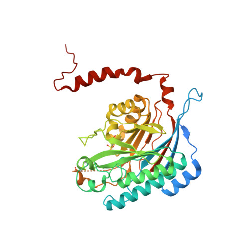
β-Ureidopropionase (βUP) catalyzes the third step of the reductive pyrimidine catabolic pathway responsible for breakdown of uracil-, thymine- and pyrimidine-based antimetabolites such as 5-fluorouracil. Nitrilase-like βUPs use a tetrad of conserved residues (Cys233, Lys196, Glu119 and Glu207) for catalysis and occur in a variety of oligomeric states. Positive co-operativity toward the substrate N -carbamoyl-β-alanine and an oligomerization-dependent mechanism of substrate activation and product inhibition have been reported for the enzymes from some species but not others. Here, the activity of recombinant human βUP is shown to be similarly regulated by substrate and product, but in a pH-dependent manner. Existing as a homodimer at pH 9, the enzyme increasingly associates to form octamers and larger oligomers with decreasing pH. Only at physiological pH is the enzyme responsive to effector binding, with N -carbamoyl-β-alanine causing association to more active higher molecular mass species, and β-alanine dissociation to inactive dimers. The parallel between the pH and ligand-induced effects suggests that protonation state changes play a crucial role in the allosteric regulation mechanism. Disruption of dimer-dimer interfaces by site-directed mutagenesis generated dimeric, inactive enzyme variants. The crystal structure of the T299C variant refined to 2.08 Å resolution revealed high structural conservation between human and fruit fly βUP, and supports the hypothesis that enzyme activation by oligomer assembly involves ordering of loop regions forming the entrance to the active site at the dimer-dimer interface, effectively positioning the catalytically important Glu207 in the active site.
Organizational Affiliation:
Department of Chemistry - BMC, Uppsala University, Box 576, SE-75123 Uppsala, Sweden.