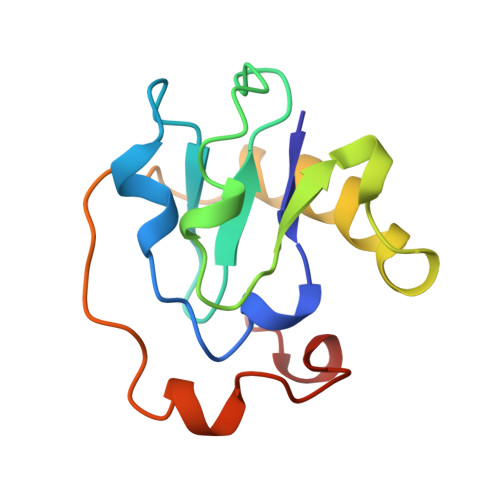
Structure of Azotobacter vinelandii 7Fe ferredoxin at 1.35 A resolution and determination of the [Fe-S] bonds with 0.01 A accuracy.
Stout, C.D., Stura, E.A., McRee, D.E.(1998) J Mol Biol 278: 629-639
- PubMed: 9600844
- DOI: https://doi.org/10.1006/jmbi.1998.1732
- Primary Citation of Related Structures:
6FD1 - PubMed Abstract:
The crystal structure of Azotobacter vinelandii ferredoxin I (FdI) at 100 K has been refined at 1.35 A resolution by full matrix block diagonal least-squares methods with anisotropic temperature factors for all non-hydrogen atoms and with hydrogen atoms included in the model. Fe-S bonds within the [3Fe-4S]+ and [4Fe-4S]2+ clusters of the protein are determined with an accuracy of at least 0.01 A. Analysis of metric parameters reveals greater variation in bonds and angles within the [3Fe-4S]+ cluster than in the [4Fe-4S]2+ cluster, whereas the opposite is true regarding the cysteine Sgamma atoms ligating to the two [Fe-S] cores. The [3Fe-4S]+ core is asymmetrically distorted by the protein matrix but relatively uniformly ligated by its three Cys ligands; in contrast the tetrahedral [4Fe-4S]2+ core is relatively symmetric but non-uniformily ligated by its four Cys ligands, three of which occur in a conserved CysxxCysxxCys residue motif. Comparison of the [3Fe-4S]+ clusters in FdI and Desulfovibrio gigas ferredoxin II, refined at 1.7 A resolution, indicates that within the limit of accuracy of the two refinements the cuboidal core is differently distorted in the two proteins. Comparison of the [3Fe-4S]+ core in FdI with the structure of a reduced [Fe3S4]o synthetic analog indicates that the protein-bound cluster displays distortions not intrinsic to the core itself. Nevertheless, both [3Fe-4S]+ and [Fe3S4]o cores have metric features consistent with expected trends due to net charge on Fe and valency of S, and both exhibit a splayed configuration with respect to their three mu2S atoms in the absence of a fourth Fe. Comparison of the [4Fe-4S]2+ cluster in FdI with the structures of [Fe4S4]2+ synthetic analogs shows that the protein bound and synthetic cubanes are very similar in geometric parameters, including the presence of tetragonal distortion in the FdI cluster common to this oxidation state.
Organizational Affiliation:
Department of Molecular Biology, The Scripps Research Institute, 10550 North Torrey Pines Road, La Jolla, CA, 92037, USA.