6EO8

Crystal structure of thrombin in complex with a novel glucose-conjugated potent inhibitor


Experimental Data Snapshot

  • Method: X-RAY DIFFRACTION
  • Resolution: 1.94 Å
  • R-Value Free: 0.248 
  • R-Value Work: 0.210 
  • R-Value Observed: 0.212 

wwPDB Validation   3D Report Full Report


Ligand Structure Quality Assessment 


This is version 1.2 of the entry. See complete history


Literature

How a beta-D-glucoside side chain enhances binding affinity to thrombin of inhibitors bearing 2-chlorothiophene as P1 moiety: crystallography, fragment deconstruction study, and evaluation of antithrombotic properties.

Belviso, B.D.Caliandro, R.de Candia, M.Zaetta, G.Lopopolo, G.Incampo, F.Colucci, M.Altomare, C.D.

(2014) J Med Chem 57: 8563-8575

  • DOI: https://doi.org/10.1021/jm5010754
  • Primary Citation of Related Structures:  
    6EO8, 6EO9

  • PubMed Abstract: 

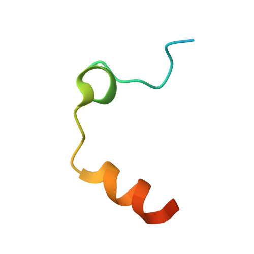
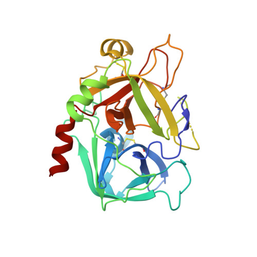
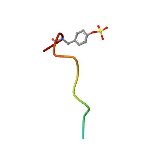
    The β-d-glucose-containing compound 3, bearing 2-chlorothiophene and 1-isopropylpiperidine moieties as binders of the S1 and S4 pockets, respectively, proved to be potent competitive inhibitor of factor Xa (fXa, Ki = 0.090 nM) and thrombin (fIIa, Ki = 100 nM). The potency of 3 increases, over the parent compound 1, against fIIa (110-fold), much more than against fXa (7-fold). Experimental deconstruction of 3 into smaller fragments revealed a binding cooperativity of the P3/P4 and propylene-linked β-d-glucose fragments, stronger in fIIa (15.5 kJ·mol(-1)) than in fXa (2.8 kJ·mol(-1)). The crystal structure of human fIIa in complex with 3 revealed a binding mode including a strong H-bond network between the glucose O1', O3', and O5' and two critical residues, namely R221a and K224, belonging to the Na(+)-binding site which may allosterically perturb the specificity sites. The potential of 3 as antithrombotic agent was supported by its ability to inhibit thrombin generation and to stimulate fibrinolysis at submicromolar concentration.


  • Organizational Affiliation

    Institute of Crystallography, Consiglio Nazionale delle Ricerche , Via Amendola 122/o, 70126 Bari, Italy.


Macromolecules
Find similar proteins by:  (by identity cutoff)  |  3D Structure
Entity ID: 1
MoleculeChains Sequence LengthOrganismDetailsImage
ProthrombinA [auth L]36Homo sapiensMutation(s): 0 
Gene Names: F2
EC: 3.4.21.5
UniProt & NIH Common Fund Data Resources
Find proteins for P00734 (Homo sapiens)
Explore P00734 
Go to UniProtKB:  P00734
PHAROS:  P00734
GTEx:  ENSG00000180210 
Entity Groups  
Sequence Clusters30% Identity50% Identity70% Identity90% Identity95% Identity100% Identity
UniProt GroupP00734
Sequence Annotations
Expand
  • Reference Sequence
Find similar proteins by:  (by identity cutoff)  |  3D Structure
Entity ID: 2
MoleculeChains Sequence LengthOrganismDetailsImage
ProthrombinB [auth H]259Homo sapiensMutation(s): 0 
Gene Names: F2
EC: 3.4.21.5
UniProt & NIH Common Fund Data Resources
Find proteins for P00734 (Homo sapiens)
Explore P00734 
Go to UniProtKB:  P00734
PHAROS:  P00734
GTEx:  ENSG00000180210 
Entity Groups  
Sequence Clusters30% Identity50% Identity70% Identity90% Identity95% Identity100% Identity
UniProt GroupP00734
Sequence Annotations
Expand
  • Reference Sequence

Find similar proteins by:  Sequence   |   3D Structure  

Entity ID: 3
MoleculeChains Sequence LengthOrganismDetailsImage
Hirudin variant-2C [auth I]12Hirudo medicinalisMutation(s): 0 
UniProt
Find proteins for P09945 (Hirudo medicinalis)
Explore P09945 
Go to UniProtKB:  P09945
Entity Groups  
Sequence Clusters30% Identity50% Identity70% Identity90% Identity95% Identity100% Identity
UniProt GroupP09945
Sequence Annotations
Expand
  • Reference Sequence
Small Molecules
Modified Residues  1 Unique
IDChains TypeFormula2D DiagramParent
TYS
Query on TYS
C [auth I]L-PEPTIDE LINKINGC9 H11 N O6 STYR
Binding Affinity Annotations 
IDSourceBinding Affinity
2FN Binding MOAD:  6EO8 Ki: 100 (nM) from 1 assay(s)
BindingDB:  6EO8 Ki: min: 0.1, max: 100 (nM) from 2 assay(s)
Experimental Data & Validation

Experimental Data

  • Method: X-RAY DIFFRACTION
  • Resolution: 1.94 Å
  • R-Value Free: 0.248 
  • R-Value Work: 0.210 
  • R-Value Observed: 0.212 
  • Space Group: C 1 2 1
Unit Cell:
Length ( Å )Angle ( ˚ )
a = 66.99α = 90
b = 71.66β = 99.7
c = 71.79γ = 90
Software Package:
Software NamePurpose
REFMACrefinement
MOSFLMdata reduction
SCALAdata scaling
Sir2014phasing

Structure Validation

View Full Validation Report


Ligand Structure Quality Assessment 


Entry History 

Deposition Data

Revision History  (Full details and data files)

  • Version 1.0: 2017-12-13
    Type: Initial release
  • Version 1.1: 2018-03-07
    Changes: Source and taxonomy
  • Version 1.2: 2019-10-16
    Changes: Data collection